Level of difficulty
Intermediate. The plexus is shallow (usually 1–3 cm), the image is normally clear and the target is easy to recognise, so the block is often among the first brachial plexus blocks that trainees learn. It is not a 'basic' block, though. The neck is crowded with vessels, the phrenic nerve lies within millimetres of C5, and the vertebral artery, the epidural space and the intrathecal space are all within reach of a needle aimed too medially or too deep.
Competence requires: confident recognition of the cervical roots and transverse processes, reliable in-plane needle-tip visualisation over a short distance, and an understanding of how volume and injection site affect diaphragmatic function. Most training programmes expect around 20–30 supervised blocks before independent practice.
Anatomy
The brachial plexus forms from the anterior (ventral) rami of C5 to T1, often with small contributions from C4 (prefixed plexus) or T2 (postfixed plexus). The rami are called roots as they leave the intervertebral foramina and run in the gutter of the transverse processes, between the anterior and posterior tubercles.
The roots pass between the anterior scalene (in front) and the middle scalene (behind), in the interscalene groove. As they descend towards the first rib they unite to form three trunks: superior (C5–C6), middle (C7) and inferior (C8–T1). The trunks then divide behind the clavicle into divisions, cords and terminal branches.
The whole complex is wrapped in a fascial sheath derived from the prevertebral layer of the deep cervical fascia. Local anaesthetic deposited within this sheath, or immediately next to it, spreads up and down the plexus.
Branches that arise in the neck (and are therefore covered by the block):
- Dorsal scapular nerve (C5): runs through or behind the middle scalene to rhomboids and levator scapulae.
- Long thoracic nerve (C5–C7): roots pierce the middle scalene and descend to serratus anterior.
- Suprascapular nerve (C5–C6): leaves the superior trunk just above the clavicle and supplies supraspinatus, infraspinatus and about 70% of the sensory supply of the shoulder joint.
- Nerve to subclavius (C5–C6), with an occasional accessory phrenic contribution.
Relations at the C6 level
- Sternocleidomastoid: superficial, its lateral (posterior) border overlying the anterior scalene.
- Internal jugular vein and common carotid artery: anteromedial, beneath sternocleidomastoid.
- Phrenic nerve (C3–C5): on the anterior surface of the anterior scalene, beneath the prevertebral fascia. At the cricoid level it is only about 2 mm from C5, and it moves further away (about 3 mm for each centimetre) as it descends. This is why the diaphragm is almost always affected by an interscalene block.
- Vertebral artery and vein: deep and medial, anterior to the C7 transverse process, entering the foramen transversarium at C6 in most people.
- Transverse cervical, suprascapular and dorsal scapular arteries: branches of the thyrocervical trunk or subclavian artery that cross the plexus at variable levels.
- Cervical sympathetic chain and recurrent laryngeal nerve: anteromedial, close enough for local anaesthetic to reach them.
- Neuraxis: the dural cuff can extend along the roots into the foramen, so a medially directed needle can reach the epidural or intrathecal space.
Variations to expect
- The C5 root (and less often C6) passes through or in front of the anterior scalene in up to a third of people. Trace it distally until it joins the groove.
- C6 often appears as two (sometimes three) hypoechoic rounds on ultrasound, because the root is already dividing into fascicles. Do not mistake these for C6 and C7.
- Vessels crossing the plexus are common, especially low in the neck, so always scan with colour Doppler.
Surface anatomy
Even when ultrasound is used, the surface landmarks tell you where to start scanning and help with the landmark technique.
- Cricoid cartilage: marks the C6 level. A horizontal line drawn laterally from it crosses the interscalene groove at the level of the C6 transverse process.
- Thyroid cartilage: roughly C4–C5, used for the higher (Meier) approach.
- Sternocleidomastoid: ask the patient to lift their head slightly off the pillow to make the clavicular head and its lateral border stand out.
- Interscalene groove: roll the fingers laterally from the posterior border of sternocleidomastoid over the belly of the anterior scalene and into the groove. A sniff or a deep breath tenses the scalenes and makes the groove easier to feel.
- C6 transverse process (Chassaignac's tubercle): the most prominent cervical transverse process, felt with firm pressure at the cricoid level.
- External jugular vein: often crosses the groove at about C6, but its position is too variable to rely on.
- Clavicle: marks the lower limit of the groove and the supraclavicular fossa where the trace-back scan begins.
Indications
- Shoulder surgery, anaesthesia or analgesia: arthroscopic subacromial decompression, rotator cuff repair, labral repair and stabilisation, capsular release, total and reverse shoulder arthroplasty, hemiarthroplasty.
- Proximal humerus surgery: fixation of proximal humeral fractures.
- Clavicle surgery: the block covers the lateral two-thirds well. For the medial clavicle and the skin over it, add a superficial cervical plexus block or rely on proximal spread.
- Acromioclavicular joint surgery.
- Upper arm surgery on the lateral side, although supraclavicular or infraclavicular blocks are usually better for the arm below the shoulder.
- Reduction of a dislocated shoulder, or physiotherapy and manipulation of a stiff shoulder.
- Pain relief for proximal humeral fractures and after shoulder trauma.
- As an awake technique (with sedation) for shoulder surgery in patients at high risk from general anaesthesia, provided they can tolerate the diaphragmatic effects.
Not a good choice for hand, wrist or forearm surgery on the ulnar side, because the inferior trunk (C8–T1) is often spared.
Nerves blocked
The block acts on the C5, C6 and usually C7 roots and the superior and middle trunks. Proximal spread commonly reaches C3–C4 (the superficial cervical plexus). Through these roots, it blocks:
| Nerve | Roots | What it supplies (relevant to surgery) |
|---|---|---|
| Suprascapular | C5–C6 | Supraspinatus, infraspinatus; most of the posterior and superior shoulder joint capsule, AC joint |
| Axillary | C5–C6 | Deltoid, teres minor; inferior joint capsule; skin over the deltoid ('regimental badge') |
| Lateral pectoral | C5–C7 | Pectoralis major; anterior joint capsule and AC joint |
| Upper and lower subscapular | C5–C6 | Subscapularis, teres major; anterior joint capsule |
| Musculocutaneous | C5–C7 | Biceps, brachialis; lateral forearm skin (lateral cutaneous nerve of forearm) |
| Radial (proximal fibres) | C5–C8 | Triceps; posterior arm |
| Supraclavicular nerves (via cervical plexus spread) | C3–C4 | Skin over the clavicle, acromion and top of the shoulder |
| Dorsal scapular, long thoracic | C5–C7 | Rhomboids, levator scapulae, serratus anterior |
Usually spared: the inferior trunk (C8–T1), so the ulnar nerve, medial cutaneous nerves of arm and forearm, and the intercostobrachial nerve (T2). For posterior portal sites or arthroscopic surgery this rarely matters. For incisions extending into the axilla, add an intercostobrachial block.
Landmark technique
Landmark and nerve-stimulator techniques are now mainly of historical and exam interest, and ultrasound guidance is the standard of care. Knowing them still helps you understand the anatomy, and you may need them when ultrasound is unavailable.
Surface anatomy, relevant structures and landmarks
- Cricoid cartilage (C6), and thyroid cartilage (C4–C5) for the Meier approach.
- Posterior (lateral) border of the clavicular head of sternocleidomastoid.
- Interscalene groove, between the anterior and middle scalene muscles.
- C6 transverse process (Chassaignac's tubercle).
- External jugular vein, which often crosses the groove at C6.
Positioning
Supine with the head on a low pillow, turned 30–45° away from the side to be blocked. The neck should be in a neutral position, not extended. The arm lies by the side, with gentle traction down towards the knee to depress the shoulder and open the groove. The operator stands at the side of the patient, at the level of the neck, so the fingers can roll across the scalenes.
Block technique
- Winnie (classic anterior) approach: at the level of the cricoid, the palpating fingers sit firmly in the interscalene groove. The needle enters between the fingers perpendicular to the skin in all planes, and is then aimed slightly caudal and slightly posterior (dorsal). As a guide, if extended, the needle would exit the back of the neck near the C7 or T1 spinous process. The plexus is usually met at 1–2.5 cm. Never advance deeper than about 2.5 cm. If you reach bone (the transverse process), walk off it rather than going deeper.
- Meier (modified lateral) approach: entry higher in the groove, at the level of the thyroid cartilage (C4–C5). The needle is directed caudally and laterally at about 30° to the skin, towards the junction of the middle and lateral thirds of the clavicle. The plexus is usually met at 3–4 cm. This trajectory runs along the groove rather than towards the spine, which lowers the risk of vertebral artery and neuraxial injection and makes it suitable for catheters.
- Borgeat (modified Winnie) and posterior (Pippa) approaches are described, but are rarely used now. The posterior approach passes through the neck muscles from behind and is uncomfortable for patients.
- If no response is found, re-enter the needle in small steps (2–3 mm) along the groove in the same plane, rather than changing the angle towards the midline.
Needle choice
22 G, 25–50 mm insulated, short-bevel (30–45°) stimulating needle. The short length is a safety feature: it limits how deep the needle can travel. For the Meier approach or a catheter, a 50 mm needle or an 18 G Tuohy catheter set.
Neurostimulation technique
Set the stimulator to 1.0 mA, 0.1 ms pulse width, 2 Hz. Advance slowly while watching the shoulder and arm. Once a plexus response appears, reduce the current step by step while making small needle adjustments, aiming to keep a clear response at 0.3–0.5 mA. If the response persists at 0.2 mA or less, the tip may be touching or inside the nerve, so withdraw slightly before injecting. If injection is painful, or if opening pressure is high (over 15 psi), stop and reposition.
Use the response you see to steer the needle:
| Response seen | Nerve stimulated | What it means and what to do |
|---|---|---|
| Deltoid (shoulder abduction) | Axillary, C5–C6 | Good. Inject. |
| Biceps (elbow flexion) | Musculocutaneous, C5–C6 | Good. Inject. |
| Pectoralis major | Lateral and medial pectoral, C5–T1 | Acceptable. |
| Triceps (elbow extension) | Radial, C7 | Acceptable. Spread will usually reach C5–C6. |
| Wrist or finger flexion or extension | C7–T1 | Acceptable, but shoulder cover may be less complete; consider moving slightly more cephalad. |
| Diaphragm (hiccup-like movement of the abdomen) | Phrenic | Needle too anterior (on the anterior scalene). Redirect posteriorly. |
| Scapular movement (rhomboids or levator scapulae) | Dorsal scapular | Needle too posterior (in the middle scalene). Redirect anteriorly. |
| Shoulder shrug (trapezius) | Accessory | Needle too posterior and superficial. Redirect anteriorly and slightly caudally. |
| Lateral chest wall (serratus anterior) | Long thoracic | Needle in the middle scalene. Redirect anteriorly. |
Choice of local anaesthetic (including volume)
With landmark technique, larger volumes were traditionally used because of the uncertainty about needle position: 20–30 mL for shoulder surgery, with 10–15 mL more through a second, more caudal injection if the forearm and hand need cover. These volumes almost always spread to the phrenic nerve, the cervical sympathetic chain and the recurrent laryngeal nerve.
Commonly used solutions: 0.5% bupivacaine or levobupivacaine, 0.5–0.75% ropivacaine, or 1.5% mepivacaine or lidocaine (with or without adrenaline 1:200,000) for shorter procedures. Inject in 3–5 mL aliquots with aspiration between each. Allow 20–30 minutes for the block to set up fully before surgery.
Ultrasound technique
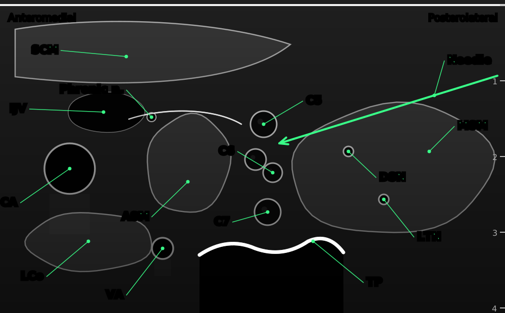

Interscalene sonoanatomy, transverse (short-axis) view at about C6–C7. Anteromedial is on the left and posterolateral on the right. The C5, C6 and C7 roots appear as round, dark structures with bright rims, stacked in the groove between the anterior and middle scalene muscles, deep to sternocleidomastoid. The needle enters in-plane through the middle scalene.
- SCM
- sternocleidomastoid
- IJV
- internal jugular vein
- CA
- common carotid artery
- ASM
- anterior scalene muscle
- MSM
- middle scalene muscle
- DSN
- dorsal scapular nerve, within the middle scalene
- LTN
- long thoracic nerve, within the middle scalene
- C5, C6, C7
- nerve roots (C6 often appears as two)
- Phrenic n.
- phrenic nerve on the anterior scalene
- LCo
- longus colli
- VA
- vertebral artery
- TP
- transverse process (posterior tubercle)
- Needle
- in-plane, lateral to medial, tip between C5 and C6
Positioning
Semi-sitting (beach-chair, about 30–45°) with the head turned slightly away and resting on a pillow. This improves the view, moves the shoulder down and is comfortable for awake patients.
Lateral position with the side to be blocked uppermost is an excellent alternative, and is our preferred position for the lateral-to-medial in-plane approach. The operator stands behind the patient, there is plenty of room for the needle behind the probe, and the patient cannot see the needle.
Place the ultrasound machine on the opposite side of the patient, directly in the operator's line of sight, so the probe, the needle and the screen are aligned. Monitoring (ECG, non-invasive blood pressure and pulse oximetry), IV access and resuscitation drugs, including lipid emulsion, should be available.
Scanning technique
- Probe orientation: transverse (short-axis) to the neck, angled slightly caudally, giving an oblique transverse view of the roots in cross-section.
- Trace-back from below (recommended for learners): place the probe in the supraclavicular fossa parallel to the clavicle. Find the subclavian artery on the first rib and the 'bunch of grapes' of trunks and divisions lateral and superficial to it. Slide the probe cranially, keeping the nerves in view, until they line up as a vertical 'traffic light' or 'snowman' stack of two or three round hypoechoic structures between the anterior and middle scalene muscles.
- From the midline: start at the cricoid with the trachea and thyroid in view. Slide laterally over the carotid artery and internal jugular vein, then on to the lateral edge of sternocleidomastoid, where the anterior scalene, the roots and the middle scalene come into view.
- Confirm the level using the transverse processes: C6 has a large anterior tubercle and a smaller posterior tubercle (a 'U' or 'two-horned' shape), with the C6 root between them. C7 has only a posterior tubercle, with the vertebral artery lying in front of it. C5 sits above C6 and often looks like a single round structure.
- Optimise the image: tilt the probe caudally and press gently. Too much pressure squeezes the internal jugular vein and the groove. Adjust depth so the plexus is in the middle of the screen, and set the focus at the level of the roots.
- Colour Doppler: scan the whole needle path with colour Doppler before needling, looking particularly for the transverse cervical, dorsal scapular and vertebral arteries.
- Scan the phrenic nerve on the surface of the anterior scalene if you plan a low-volume, phrenic-sparing technique. It appears as a small hypoechoic dot or oval under the prevertebral fascia.
Structures to identify
- Sternocleidomastoid (superficial)
- Internal jugular vein and common carotid artery (anteromedial)
- Anterior scalene muscle
- Middle scalene muscle
- C5 root (sometimes within the anterior scalene)
- C6 root, often appearing as two rounds
- C7 root
- Superior trunk (lower in the neck, where C5 and C6 join)
- Phrenic nerve on the anterior surface of the anterior scalene
- Vertebral artery (anterior to the C7 transverse process)
- C6 and C7 transverse processes, with their tubercles
- Dorsal scapular and long thoracic nerves within the middle scalene
- Transverse cervical or other arteries crossing the groove (with colour Doppler)
Block performance


Position / ergonomics
Semi-sitting with the head turned away, or lateral with the operative side up. Stand at the head of the bed or behind the patient so that the needle enters from the posterolateral side. Raise the bed so that your forearm can rest on the patient, and keep your probe hand steady by resting its little finger on the patient's skin. Keep the screen directly in front of you, beyond the patient, so that you look at the probe, the needle and the screen in one line.
Use a full aseptic technique: hand hygiene, sterile gloves, skin preparation with chlorhexidine in alcohol (allowed to dry), a sterile probe cover and sterile gel. Perform the 'Stop Before You Block' check to confirm the side immediately before needle insertion.
Probe
High-frequency linear array probe, 8–15 MHz (or 6–13 MHz). A small-footprint ('hockey stick') linear probe helps in short or thick necks, and in children.
Settings
Nerve or small-parts preset. Set the single focal zone at the depth of the roots. Use enough gain so the muscle looks mid-grey and the roots look clearly black with a bright rim. Use harmonic imaging and compound imaging if available, as these improve the outline of the roots. Have colour Doppler ready, with the velocity scale set low (about 15–25 cm/s) to detect small arteries.
Depth
Start at 3–4 cm. The roots usually lie 1–2 cm beneath the skin (up to 3 cm in larger patients). Reduce the depth so that the plexus sits in the middle of the screen, with the transverse process visible at the bottom.
Needle size
22 G, 50 mm short-bevel, echogenic, insulated stimulating needle. A 50 mm needle is long enough for the lateral in-plane approach in most adults; avoid longer needles. Use an 18 G Tuohy or catheter-over-needle system for continuous techniques.
Needling technique
- In-plane, lateral to medial (posterolateral approach): the standard approach. Insert the needle 1–2 cm from the lateral end of the probe and pass it through the middle scalene into the groove. This route keeps the needle away from the phrenic nerve, the carotid artery and the internal jugular vein. Look for the dorsal scapular and long thoracic nerves in the middle scalene, and redirect if they lie in your path.
- A faint 'pop' is often felt as the needle passes through the fascia of the middle scalene into the interscalene groove.
- In-plane, medial to lateral: possible, but the needle crosses the anterior scalene and passes close to the phrenic nerve, the carotid artery and the internal jugular vein. Avoid it where possible.
- Out-of-plane (cranial to caudal): the probe sits transverse and the needle enters just above it, aimed caudally along the groove. This route has a short path and is useful for catheters, but the tip is harder to follow. Use hydrodissection with small volumes to find it.
- Before injecting, aspirate. Then inject 0.5–1 mL to confirm that the tip is in the right plane. Spread should separate the roots from the scalene muscles rather than distend a root.
- Inject slowly in 3–5 mL increments, aspirating between them, watching the spread and asking the patient about pain or paraesthesia.
Optimal needle-tip position
Within the interscalene groove, either between C5 and C6, or just lateral (posterior) to C5–C6, close to the nerves but outside their epineurium. The ideal spread surrounds C5 and C6 like a halo, and can be seen tracking up and down the groove when you slide the probe.
Avoid placing the tip between the fascicles of a single root (for example, between the two 'rounds' of a divided C6), because this is likely to be intraneural. The roots in the neck have more nerve tissue and less protective connective tissue than peripheral nerves, so intraneural injection here is more harmful.
Extra-sheath option: depositing the local anaesthetic just outside the sheath, within the middle scalene a few millimetres from C5, gives similar analgesia, may lower the risk of nerve injury and may slightly reduce phrenic involvement, at the cost of a slightly slower onset.
Drug choice
| Goal | Drug | Typical duration |
|---|---|---|
| Surgical anaesthesia (long) | 0.5% bupivacaine or levobupivacaine; 0.75% ropivacaine | Anaesthesia 4–8 h; analgesia 12–18 h |
| Analgesia (with GA) | 0.25% bupivacaine or levobupivacaine; 0.375–0.5% ropivacaine | 8–16 h |
| Short procedures or day surgery | 1.5% mepivacaine; 1.5–2% lidocaine (± adrenaline) | 2–5 h |
| Faster onset with long duration | Mixture of 2% lidocaine and 0.5% bupivacaine (1:1) | Faster onset; shorter duration than bupivacaine alone |
Adjuvants: dexamethasone prolongs analgesia by several hours. IV dexamethasone (for example 0.1–0.2 mg/kg, up to 8 mg) is nearly as effective as the perineural route, and avoids off-label perineural use and the risk of crystallisation with some local anaesthetics. Most clinicians now prefer the IV route. Perineural dexmedetomidine also prolongs the block, but can cause sedation, bradycardia and hypotension.
Liposomal bupivacaine (where available) can extend analgesia to about 72 hours.
Maximum doses: keep within the recommended safe dose for the patient's weight (for example bupivacaine 2 mg/kg, ropivacaine 3 mg/kg), especially if other blocks or wound infiltration are planned.
Avoid long-acting high-concentration solutions in patients who could not tolerate a 25–30% fall in their vital capacity.
Volume
Analgesia: 5–10 mL is enough for most shoulder surgery when combined with general anaesthesia. Studies show minimum effective volumes of about 5 mL or less with ultrasound guidance.
Surgical anaesthesia (awake surgery): 10–15 mL, sometimes up to 20 mL.
Lower volumes reduce, but do not reliably prevent, hemidiaphragmatic paresis. Volumes above 15–20 mL rarely improve analgesia, and increase spread to the phrenic, recurrent laryngeal and sympathetic nerves.
Optimal current if nerve stimulation is used
With ultrasound guidance, nerve stimulation is used as a safety monitor rather than to find the nerve. Set it to 0.5 mA (0.1 ms, 2 Hz). A motor response at or below 0.5 mA means the tip is very close to, or in contact with, the nerve, so stop advancing and withdraw slightly. A response below 0.2 mA suggests intraneural placement.
With the stimulator as the main guide (no ultrasound), aim for a clear response between 0.3 and 0.5 mA before injecting.
Muscle responses acceptable
- Deltoid contraction (shoulder abduction): axillary nerve, C5–C6. The best response for shoulder surgery.
- Biceps contraction (elbow flexion): musculocutaneous nerve, C5–C6.
- Pectoralis major contraction: pectoral nerves.
- Triceps contraction (elbow extension): radial nerve, C7. Acceptable.
- Not acceptable: diaphragm (phrenic), scapular or rhomboid movement (dorsal scapular), trapezius (accessory) or serratus anterior (long thoracic). See the table in the landmark section for how to redirect.
Testing block success
Test at 5, 10 and 20 minutes. Motor block usually appears before sensory block. Most blocks are ready for surgery by 15–30 minutes with long-acting drugs, and sooner with lidocaine or mepivacaine.
- Motor (the 'deltoid sign'): the patient cannot abduct the arm against resistance (deltoid, C5–C6).
- Motor: weak or absent elbow flexion (biceps, C5–C6), and often weak elbow extension (triceps, C7).
- Sensory: loss of cold sensation or pinprick over the deltoid ('regimental badge' area, axillary nerve, C5), the lateral arm and the lateral forearm (C6).
- Sensory: over the clavicle and acromion (supraclavicular nerves, C3–C4) if cervical plexus spread has occurred.
- Sparing of the medial arm, forearm and little finger (C8–T1) is normal and expected.
- Hoarseness, Horner syndrome or mild breathlessness at this stage are signs of spread, not failure, and show the block is working.
Continuous techniques
Indication
Major shoulder surgery with severe pain expected beyond the 12–18 hours of a single injection: total or reverse shoulder arthroplasty, open rotator cuff repair, proximal humeral fracture fixation, and capsular release followed by early intensive physiotherapy. It is also used for patients who do not tolerate opioids, and for prolonged pain relief after severe trauma.
Recent trials show that, with good multimodal analgesia (including dexamethasone), a single injection may give similar overall results to a catheter for many patients. Reserve catheters for patients most likely to benefit, and where there is a service to manage them.
Kit
- Ultrasound machine with a linear probe and a long sterile probe cover
- Full sterile set: gown, gloves, drape, chlorhexidine in alcohol
- Continuous nerve block set: 18 G Tuohy needle (50 mm) with a 20 G multi-orifice catheter, or a catheter-over-needle system
- Local anaesthetic for skin and for the bolus, and saline for hydrodissection
- Skin glue, clear occlusive dressing and catheter fixation device
- Labelled infusion pump (elastomeric or electronic, ideally with a patient-controlled bolus function) and a 'regional block' label for the line
Catheter
- Place the catheter through the middle scalene using a lateral-to-medial in-plane approach, or through an out-of-plane approach along the groove. Passing through muscle helps anchor the catheter and reduces dislodgement.
- Open the space with saline or local anaesthetic first, then advance the catheter 2–3 cm beyond the needle tip. Do not advance it further, as long catheters can coil away from the plexus or migrate towards the neuraxis.
- Confirm the position by injecting through the catheter while watching the spread on ultrasound. Colour Doppler can help show where the injectate goes.
- Seal the entry site with skin glue to reduce leakage, apply a clear occlusive dressing, and tunnel the catheter if it is to stay in for more than a couple of days.
Regime
Initial bolus: 5–10 mL of 0.2–0.5% ropivacaine or 0.25% levobupivacaine through the needle or catheter.
Infusion: 0.2% ropivacaine or 0.1–0.125% levobupivacaine at 4–6 mL/h. Combining a lower background rate (for example 2–5 mL/h) with patient-controlled boluses of 4–5 mL (lockout 30–60 minutes) uses less drug and gives similar or better pain relief.
Duration: usually 48–72 hours. Review daily for pain scores, catheter site, leakage, motor block, breathing and signs of local anaesthetic toxicity.
Breakthrough pain: give a bolus, check the catheter position with ultrasound, and consider increasing the rate. If the catheter has dislodged, replace or remove it and use oral analgesia.
Patients going home with a catheter need written instructions, a 24-hour contact number and a plan for removal.
A continuous infusion keeps the hemidiaphragm paralysed for as long as it runs, so be cautious in patients with poor respiratory reserve.
Video demonstrations
Videos play from YouTube. They load only when you press play.
Contraindications to the block
Absolute
- Patient refusal or lack of consent
- Infection at the injection site
- True allergy to local anaesthetic
- Known contralateral phrenic nerve palsy or a paralysed contralateral hemidiaphragm
- Known contralateral recurrent laryngeal nerve palsy (risk of complete airway obstruction)
- Bilateral interscalene block (never perform both sides)
Relative
- Severe respiratory disease: COPD, restrictive lung disease, obesity hypoventilation, severe obstructive sleep apnoea, or any patient who could not tolerate a 25–30% fall in vital capacity
- Morbid obesity, where a reduced diaphragm movement is poorly tolerated
- Coagulopathy or anticoagulation: the site is compressible, but a haematoma in the neck can compress the airway (follow ASRA/ESRA guidance)
- Pre-existing neurological deficit in the arm, or neuropathy (document carefully before the block)
- Contralateral pneumonectomy or lung resection
- Previous neck surgery or radiotherapy distorting the anatomy
- Patient unable to cooperate or keep still, or unable to report pain during injection
- Children under general anaesthesia (performed only by experienced operators, if at all)
Side effects and complications
Expected side effects
- Ipsilateral hemidiaphragmatic paresis: close to 100% with 20 mL or more, and still about 30–60% with 5 mL. Vital capacity falls by about 25–30%. Most healthy patients do not notice, but some feel short of breath.
- Horner syndrome (ptosis, miosis, anhidrosis, enophthalmos) from spread to the cervical sympathetic chain: common and harmless.
- Hoarseness from recurrent laryngeal nerve block: in about 10–20% of patients.
- Tinnitus or a feeling of fullness in the ear, and mild difficulty swallowing.
- Numbness of the skin over the neck and clavicle from cervical plexus spread.
- Rebound pain when the block wears off, usually in the first night.
Complications
- Vertebral artery injection: very small doses (1–2 mL) can cause immediate seizures.
- Epidural, subdural or intrathecal injection: from a medially directed needle or injection along a dural sleeve, causing a high or total spinal block.
- Local anaesthetic systemic toxicity (LAST): from intravascular injection or excessive dose.
- Nerve injury: transient postoperative symptoms in up to about 10% at 10 days, falling to under 1% at 6 months. Permanent injury is rare (about 1–4 per 10,000). Surgery, positioning and tourniquets also contribute.
- Pneumothorax: uncommon at the interscalene level, but possible if the needle is directed caudally and deep, or if the block is performed low in the neck.
- Haematoma and vascular puncture of the carotid artery or internal jugular vein.
- Bezold–Jarisch reflex: sudden hypotension and bradycardia in the sitting position, especially with adrenaline-containing solutions.
- Persistent phrenic nerve palsy: rare, but reported.
- Respiratory failure in patients with poor reserve, or contralateral phrenic palsy.
- Infection, particularly with catheters left in place for several days.
Pearls of wisdom
- Trace back from the supraclavicular fossa if the groove is hard to read. The plexus is easiest to recognise next to the subclavian artery.
- Scan with colour Doppler every time. A transverse cervical artery can lie right on the plexus.
- Aim between C5 and C6, or just lateral to them, never into the middle of a root. Remember that the two 'rounds' of a divided C6 are one root.
- Count the transverse processes to confirm the level: C7 has only a posterior tubercle, with the vertebral artery in front of it.
- Use the lateral approach through the middle scalene. It keeps the needle away from the phrenic nerve and the great vessels, and the muscle anchors any catheter.
- Low volume is enough for analgesia. 5–10 mL works for most arthroscopic surgery when combined with general anaesthesia.
- Inject with low pressure. If you feel resistance or the patient reports severe pain, stop. High opening pressure (over 15 psi) suggests needle–nerve contact.
- For poor respiratory reserve, consider a phrenic-sparing alternative: superior trunk block, anterior suprascapular block, suprascapular plus axillary nerve blocks, or the anterior glenoid block.
- For clavicle surgery, the interscalene block on its own often gives enough cover. Add a superficial cervical plexus block if the incision is medial or analgesia is incomplete.
- Tell the patient in advance that a droopy eyelid, a hoarse voice or mild breathlessness are expected and will wear off.
- Plan for rebound pain: give the first doses of regular oral analgesia before the block wears off, typically the first evening.
- Beach-chair hypotension: be ready to treat sudden bradycardia and hypotension once the patient is sat up.
Review of literature
Diaphragmatic function
- Landmark interscalene block with large volumes caused ipsilateral hemidiaphragmatic paresis in every patient studied, confirmed with ultrasound, with falls in FVC and FEV1 of about 25–30% (Urmey 1991).
- Reducing the volume from 20 to 5 mL under ultrasound guidance reduced diaphragmatic paresis and the fall in lung function, with similar analgesia (Riazi 2008). Low-dose injection away from C5, for example at C7, also reduced paresis (Renes 2009).
- A 2025 meta-analysis of 28 randomised trials found that low-volume interscalene block reduced complete hemidiaphragmatic paralysis compared with conventional volumes (risk ratio 0.62), and that the extrafascial technique and lower concentrations also reduced it. The evidence was strongest for the extrafascial technique (Oliver-Fornies 2025).
- No low-volume interscalene technique reliably avoids the phrenic nerve. The review by El-Boghdadly et al (2017) summarises the anatomy and the alternatives.
Volume and injection site
- Ultrasound guidance reduced the minimum effective volume compared with nerve stimulation alone (McNaught 2011). Minimum effective volumes of about 5 mL or less have been reported for shoulder analgesia (Gautier 2011; Falcão 2013).
- Injecting outside the sheath, lateral to C5–C6, gave analgesia similar to injecting inside it (Spence 2011). A needle-tip distance of up to about 4 mm from the plexus was still effective (Albrecht 2014).
Superior trunk block as an alternative
- Meta-analyses of randomised trials show that the superior trunk block causes far less complete hemidiaphragmatic paralysis than interscalene block (risk ratio about 0.07–0.10), and less Horner syndrome and breathlessness. Pain scores, opioid use and block duration were similar (Amaral 2023, 4 trials; da Cunha 2026, 8 trials). Not every analysis was conclusive (Oliver-Fornies 2025).
Adjuvants
- Both perineural and IV dexamethasone prolong the block. A 2024 meta-analysis found that perineural dexamethasone extended analgesia by only about 2 hours more than the IV route, with no difference in pain scores or opioid use. Most authors now recommend the IV route (Albrecht 2024).
Single injection versus catheter
- In the setting of good multimodal analgesia, adding a continuous infusion to a single injection did not improve pain control or opioid use after shoulder surgery (Rhyner 2024). Catheters are therefore increasingly kept for selected patients.
Safety
- Ultrasound guidance reduces the volume needed and the rate of vascular puncture. Its effect on rare outcomes, such as permanent nerve injury, is harder to prove, but large registries show these remain rare.
Comparison with other blocks
The interscalene block remains the reference standard for shoulder analgesia. The alternatives below aim to keep most of its benefit while sparing the diaphragm.
| Block | Analgesia for shoulder | Diaphragm effect | Notes |
|---|---|---|---|
| Interscalene | Reference standard | Near 100% at standard volume; reduced, not removed, with low volume | Most reliable and quickest. Accept the phrenic block or reduce the volume. |
| Superior trunk | Similar to interscalene (meta-analyses) | Much lower: complete paralysis cut by about 90% | Performed just distal to where C5 and C6 join, below the phrenic nerve's closest point (Burckett-St Laurent 2014). |
| Suprascapular plus axillary | Slightly less in the first hours | Minimal | Good choice for respiratory disease (Hussain 2017). Two injections. |
| Anterior suprascapular | Slightly less than interscalene | Lower | Phrenic-sparing alternative in the supraclavicular fossa; analgesia non-inferior in a multicentre trial (Abdallah 2020). |
| Anterior glenoid block (with or without suprascapular) | Promising; evidence limited | Minimal | Newer block that targets the articular branches to the front of the joint (2024 studies). |
| Supraclavicular | Good for shoulder when spread is high | About 30–50% | Small risk of pneumothorax. Covers the whole arm. |
| Costoclavicular | Inconsistent for shoulder | Low | Mainly for elbow, forearm and hand. |
| Periarticular infiltration (by surgeon) | Shorter and less effective | None | Useful when a block is contraindicated. |
References
- Urmey WF, Talts KH, Sharrock NE. One hundred percent incidence of hemidiaphragmatic paresis associated with interscalene brachial plexus anesthesia as diagnosed by ultrasonography. Anesth Analg 1991;72:498–503. PubMed
- Riazi S, Carmichael N, Awad I, Holtby RM, McCartney CJL. Effect of local anaesthetic volume (20 vs 5 ml) on the efficacy and respiratory consequences of ultrasound-guided interscalene brachial plexus block. Br J Anaesth 2008;101:549–556. PubMed
- Renes SH, Rettig HC, Gielen MJ, Wilder-Smith OH, van Geffen GJ. Ultrasound-guided low-dose interscalene brachial plexus block reduces the incidence of hemidiaphragmatic paresis. Reg Anesth Pain Med 2009;34:498–502. PubMed
- El-Boghdadly K, Chin KJ, Chan VWS. Phrenic nerve palsy and regional anesthesia for shoulder surgery: anatomical, physiologic, and clinical considerations. Anesthesiology 2017;127:173–191. PubMed
- McNaught A, Shastri U, Carmichael N, et al. Ultrasound reduces the minimum effective local anaesthetic volume compared with peripheral nerve stimulation for interscalene block. Br J Anaesth 2011;106:124–130. PubMed
- Gautier P, Vandepitte C, Ramquet C, et al. The minimum effective anesthetic volume of 0.75% ropivacaine in ultrasound-guided interscalene brachial plexus block. Anesth Analg 2011;113:951–955. PubMed
- Falcão LF, Perez MV, de Castro I, et al. Minimum effective volume of 0.5% bupivacaine with epinephrine in ultrasound-guided interscalene brachial plexus block. Br J Anaesth 2013;110:450–455. PubMed
- Spence BC, Beach ML, Gallagher JD, Sites BD. Ultrasound-guided interscalene blocks: understanding where to inject the local anaesthetic. Anaesthesia 2011;66:509–514. PubMed
- Albrecht E, Kirkham KR, Taffé P, et al. The maximum effective needle-to-nerve distance for ultrasound-guided interscalene block: an exploratory study. Reg Anesth Pain Med 2014;39:56–60. PubMed
- Albrecht E, Renard Y, Desai N. Intravenous versus perineural dexamethasone to prolong analgesia after interscalene brachial plexus block: a systematic review with meta-analysis and trial sequential analysis. Br J Anaesth 2024;133:135–145. PubMed
- Rhyner P, Cachemaille M, Goetti P, et al. Single-bolus injection of local anesthetic, with or without continuous infusion, for interscalene brachial plexus block in the setting of multimodal analgesia: a randomized controlled unblinded trial. Reg Anesth Pain Med 2024;49:313–319. PubMed
- Burckett-St Laurent D, Chan V, Chin KJ. Refining the ultrasound-guided interscalene brachial plexus block: the superior trunk approach. Can J Anaesth 2014;61:1098–1102. PubMed
- Hussain N, Goldar G, Ragina N, et al. Suprascapular and interscalene nerve block for shoulder surgery: a systematic review and meta-analysis. Anesthesiology 2017;127:998–1013. PubMed
- Abdallah FW, Wijeysundera DN, Laupacis A, et al. Subomohyoid anterior suprascapular block versus interscalene block for arthroscopic shoulder surgery: a multicenter randomized trial. Anesthesiology 2020;132:839–853. PubMed
- Hadzic A (ed). Hadzic's Peripheral Nerve Blocks and Anatomy for Ultrasound-Guided Regional Anesthesia. McGraw-Hill.
- Farag E, Brown DL. Interscalene block. In: Brown's Atlas of Regional Anesthesia, 5th ed. Elsevier, 2017.
- Amaral S, Arsky Lombardi R, Medeiros H, Nogueira A, Gadsden J. Superior trunk block is an effective phrenic-sparing alternative to interscalene block for shoulder arthroscopy: a systematic review and meta-analysis. Cureus 2023;15:e48217. doi:10.7759/cureus.48217
- Oliver-Fornies P, Aragon-Benedi C, Gomez Gomez R, et al. Hemidiaphragmatic paralysis after ultrasound-guided brachial plexus blocks for shoulder surgery: a systematic review and meta-analysis of randomized clinical trials. J Clin Anesth 2025;105:111874. doi:10.1016/j.jclinane.2025.111874
- da Cunha DF, de Carvalho JPA, de Olinda Ribeiro B, et al. Incidence of hemidiaphragmatic paralysis in superior trunk versus interscalene block upper limb surgeries: a systematic review, meta-analysis, and trial sequential analysis. Anesth Pain Med (Seoul) 2026. doi:10.17085/apm.25415
- Phillips S, et al (eds). Regional Anaesthesia: A Guide to Clinical Exams. Springer Nature, 2026.