1Learning objectives
- Describe how each class of antithrombotic drug works and how long its effect lasts.
- Find the interval before a block, and before restarting the drug, for a given drug, dose and renal function.
- Explain how ASRA and ESAIC/ESRA guidance differ, and why.
- Classify a block as deep or superficial, compressible or not, and apply the right rule.
- Plan catheter insertion and removal around anticoagulant doses.
- Choose and interpret coagulation tests and drug levels.
- Make and document a risk–benefit decision with the surgical and medical teams.
2Overview
Always check the current guideline and local policy for the exact drug, dose, renal function and block. The tables here are for learning and comparison, not a prescription.
Bleeding into the spinal canal after neuraxial anaesthesia is rare but can cause permanent paralysis; bleeding after deep peripheral blocks can cause nerve compression, large haematomas and transfusion. The risk rises with antiplatelet and anticoagulant drugs. Stopping those drugs has its own risk: stroke, coronary stent thrombosis, pulmonary embolism.
This module supports that assessment for the individual patient. There is no single safe interval for all drugs, doses, block techniques and renal dysfunction, and the two main guidelines do not always agree. Where they differ, both are shown, to explain the basis of local hospital policy.
3Pharmacology of antithrombotic drugs
Drug classes, renal function and tests
Antiplatelet agents
Aspirin and NSAIDs inhibit cyclo-oxygenase; alone they add little neuraxial risk. P2Y12 inhibitors (clopidogrel, prasugrel, ticagrelor) need days to wear off because platelets must be replaced (clopidogrel, prasugrel) or the drug must clear (ticagrelor). Dual antiplatelet therapy after a coronary stent should not be stopped without cardiology advice.
Heparins
Unfractionated heparin (UFH) has a short half-life and is monitored with aPTT, anti-Xa or ACT; protamine reverses it. Low molecular weight heparins (LMWH) last longer, are cleared by the kidneys, are monitored with anti-Xa, and are only partly reversed by protamine. Prophylactic and therapeutic doses have very different intervals.
Warfarin
Vitamin K antagonists reduce factors II, VII, IX and X. The effect fades over about 5 days after stopping; the INR measures it. Reversal: vitamin K and prothrombin complex concentrate. Early after stopping, the INR may look normal while factors II and X are still low.
DOACs
Direct oral anticoagulants inhibit factor Xa (apixaban, rivaroxaban, edoxaban) or thrombin (dabigatran). They act within hours, have half-lives of about 5–17 h, and depend on the kidneys to varying degrees (dabigatran most). Routine tests (INR, aPTT) do not reliably show their effect; drug-specific levels do.
Other anticoagulants
Fondaparinux (indirect factor Xa inhibitor) has a long half-life and is renally cleared. Parenteral direct thrombin inhibitors (argatroban, bivalirudin) are short-acting but neuraxial techniques are not advised by ASRA. Thrombolytics break down clot and need the longest caution.
Renal impairment
LMWH, fondaparinux and dabigatran accumulate when creatinine clearance falls; Xa inhibitors less so. Both guidelines lengthen intervals or ask for a level in renal impairment, but by different rules (see the comparison table). Calculate creatinine clearance (Cockcroft–Gault) with an up-to-date creatinine.
Coagulation testing
INR for warfarin; aPTT, anti-Xa or ACT for unfractionated heparin; anti-Xa for LMWH and (calibrated) for fondaparinux and Xa inhibitors; dilute thrombin time or ecarin-based tests for dabigatran; platelet count for thrombocytopenia. A normal INR or aPTT does not exclude a DOAC effect. In obstetric thrombocytopenia, an interdisciplinary consensus supports neuraxial procedures at platelet counts of 70 × 10⁹/L or more, without other risk factors (Bauer 2021).
4Bleeding risk and block site
Deep versus superficial blocks
Both guidelines treat deep blocks like neuraxial blocks for timing. The test is not just depth in centimetres, but whether bleeding would be seen early and could be stopped by pressure, and what harm it could do.
- Neuraxial: spinal, epidural, combined spinal–epidural, caudal. Bleeding is hidden and can compress the spinal cord.
- Deep, non-compressible: ASRA lists lumbar plexus, paravertebral, infraclavicular and stellate ganglion among them. Bleeding can be retroperitoneal, pleural or in the neck.
- Superficial, compressible: many limb and some fascial plane blocks. ESAIC/ESRA: neuraxial intervals do not apply, after a risk–benefit check; its Table 2 classifies each block.
- Uncertain: some plane blocks (for example erector spinae, quadratus lumborum approaches) sit between the two. Check the guideline tables and local policy; when unsure, treat as deep.
Compressibility
- Is there a firm structure (bone, a muscle against bone) to press against?
- Is the vessel near the needle path large or small, and is it visible on ultrasound and colour Doppler?
- Would bleeding be visible early, or hidden in a body cavity?
- What would a haematoma compress: a nerve in a tight compartment, the airway, the spinal cord?
 Illustration
Axillary block: superficial, with the artery easily compressed against the humerus. Axillary block
 Ultrasound
 Illustration
Infraclavicular block: a deep site under two muscles near the axillary vessels (ASRA lists it as deep). Infraclavicular block
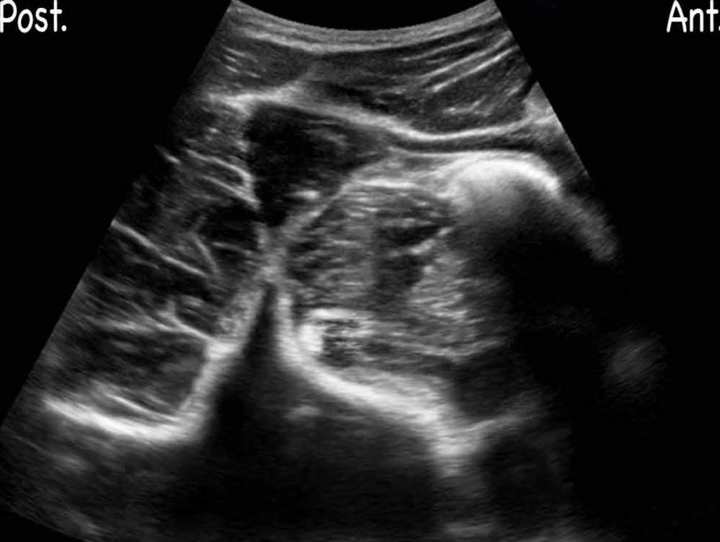 Ultrasound
 Illustration
Lumbar plexus block: deep, in psoas, with retroperitoneal bleeding risk. Lumbar plexus block
The role of ultrasound
Ultrasound allows many vessels to be identified and avoided, but neither guideline shortens intervals because of it (ESAIC/ESRA states this explicitly). Use it to lower the chance of a bloody tap, not to justify an earlier block.
- Pre-scan the whole needle path with colour Doppler, at low velocity scale for small vessels.
- Choose a path that avoids vessels even if it is longer.
- Watch for haematoma: a new dark area that grows, or loss of the normal planes after a vascular puncture.
- A bloody tap changes the plan: both guidelines advise delaying the next anticoagulant dose (ASRA: delay the first LMWH dose by 24 h).
5Timing of interruption and resumption
Timing of medication interruption and resumption
Two times matter for every drug: the gap from the last dose to the needle (or catheter removal), and the gap from the needle (or catheter removal) to the next dose. The table compares the 'before' intervals in the ESAIC/ESRA and ASRA guidelines, with the UK 2013 guideline (Table 1, values unchanged) alongside. Type a drug to filter.
| Drug | ESAIC/ESRA 2022: stop before | ASRA 2025: stop before | AAGBI/OAA/RA-UK 2013: block after | Agreement and differences |
|---|---|---|---|---|
| Aspirin (low dose) | No interval after a risk–benefit check; a single-shot spinal preferred to an epidural | No interval (aspirin and other NSAIDs) | No additional precautions | All three: no interval. ESAIC/ESRA adds a preference for single-shot neuraxial |
| Aspirin (high dose) | 3 days (normal platelet count) to 7 days | No interval | No additional precautions | Differs: only ESAIC/ESRA sets an interval for high-dose aspirin; ASRA and UK set none |
| Clopidogrel | 5–7 days | 5–7 days | 7 days | Similar: 5–7 days (UK 7 days); restart rules differ in detail |
| Prasugrel | 7 days | 7–10 days | 7 days | Differs: ASRA allows up to 10 days; ESAIC/ESRA and UK 7 days |
| Ticagrelor | 5 days | 5 days | 5 days | Same in all three (5 days) |
| UFH, low-dose SC | 4 h | 4–6 h, or normal coagulation tests | 4 h or normal APTTR | Similar: about 4 h; ASRA and UK also accept normal coagulation tests |
| UFH, IV infusion | 6 h, with normal aPTT, anti-Xa or ACT | 4–6 h and normal coagulation tests | 4 h or normal APTTR | Similar: 4–6 h; all three use normal coagulation tests |
| LMWH, prophylactic | 12 h (24 h if CrCl below 30 mL/min) | 12 h (check anti-Xa ≤0.1 IU/mL if sooner) | 12 h | Same 12 h in all three; only ESAIC/ESRA extends it in renal impairment |
| LMWH, therapeutic | 24 h (48 h if CrCl below 30), or anti-Xa ≤0.1 IU/mL | 24 h (anti-Xa ≤0.1 IU/mL, especially over 75 years or CrCl ≤30) | 24 h | Same 24 h in all three; renal approach differs (ESAIC/ESRA longer wait, ASRA anti-Xa level, UK assumes normal renal function) |
| Fondaparinux, prophylactic | 36 h (72 h if CrCl below 50) | 36 h (younger) to 42 h (elderly); 58 h if CrCl 30–50; avoid if CrCl below 30 | 36–42 h (consider anti-Xa levels) | Similar base (36–42 h); renal and age adjustments differ (ESAIC/ESRA and ASRA) |
| Warfarin | About 5 days and INR normal (INR below 1.5 may be acceptable after risk–benefit check) | 5 days and normal INR | INR ≤ 1.4 | Similar: about 5 days; UK threshold INR ≤ 1.4, ESAIC/ESRA below 1.5 after risk–benefit check |
| Dabigatran | Low dose 48 h, high dose 72 h. CrCl below 50: level below 30 ng/mL or normal thrombin time | Low 48 h, high 72 h (120 h if CrCl 30–49). CrCl below 30: only with a level below 30 ng/mL | 48 h (CrCl > 80); 72 h (CrCl 50–80); 96 h (CrCl 30–50) | Differs in renal impairment: longer fixed times (ASRA, UK by creatinine clearance) versus a drug level (ESAIC/ESRA) |
| Rivaroxaban | Low 24 h (30 h if CrCl below 30). High 72 h, or level below 30 ng/mL if CrCl below 30 | Low 24 h (30 h if CrCl below 30). High 72 h, or level below 30 ng/mL | Prophylaxis 18 h; treatment 48 h (CrCl > 30) | Differs: UK intervals are shorter (18 h prophylaxis, 48 h treatment) than ESAIC/ESRA and ASRA (24 h and 72 h) |
| Apixaban | Low 36 h. High 72 h, or level below 30 ng/mL | Low 36 h. High 72 h, or level below 30 ng/mL | Prophylaxis 24–48 h (treatment dose not listed) | Differs: UK prophylaxis 24–48 h versus 36 h; UK gives no treatment-dose interval |
| Edoxaban | Low 24 h (30 h if CrCl below 30). High 72 h | High 72 h, or level below 30 ng/mL. No approved low dose | Not listed (the guideline predates edoxaban) | ESAIC/ESRA and ASRA: same therapeutic interval; ASRA lists no low dose; not in the UK guideline |
Restart times and every drug in each guideline: Anticoagulants and blocks (full ESAIC/ESRA and ASRA tables).
How the three guidelines compare in approach
| Question | ESAIC/ESRA 2022 | ASRA 2025 (5th edition) | AAGBI/OAA/RA-UK 2013 |
|---|---|---|---|
| Which blocks follow neuraxial timing? | Deep, non-compressible peripheral blocks. Superficial, compressible blocks may be done on antithrombotic drugs after a risk–benefit check; the guideline's Table 2 classifies blocks. | Deep plexus and deep peripheral blocks (for example lumbar plexus, paravertebral, infraclavicular, stellate ganglion). Other blocks: judge compressibility, vascularity and the consequence of bleeding. | Table 2 ranks blocks on a continuum of relative risk: epidural catheter (highest), single-shot epidural, spinal, paravertebral, deep, superficial perivascular, fascial and superficial blocks, then local infiltration (normal risk). |
| Method | GRADE with Delphi consensus; certainty of evidence low (GRADE C) throughout. | Evidence-based review and expert consensus; 5th edition. | Consensus of an expert Working Party (AAGBI, OAA, RA-UK), based on existing guidance and drug pharmacology; intervals derived from time to peak effect plus two elimination half-lives. |
| Renal impairment | Adjusts many intervals by creatinine clearance; uses drug levels for some DOACs. | Adjusts some intervals (dabigatran, fondaparinux, LMWH) and accepts levels or anti-Xa. | Intervals assume normal renal function except where stated (dabigatran and rivaroxaban by creatinine clearance). |
| Drug levels | Accepts measured levels or anti-Xa to shorten some waits. | Accepts drug level below 30 ng/mL or anti-Xa ≤0.1 IU/mL as alternatives. | Suggests anti-Xa levels for danaparoid and fondaparinux, and normal APTTR for heparins. |
| Ultrasound and risk scores | Do not shorten the intervals. | Ultrasound is not discussed as a reason to shorten intervals. | Ultrasound in experienced hands may reduce vascular puncture and make blocks such as supraclavicular safer when coagulation is altered. |
| Reversal for a routine block | Specific reversal (vitamin K antagonists, dabigatran) may change intervals. | Do not use DOAC reversal drugs just to allow a routine block. | No specific recommendation on reversal to enable a routine block. |
Common to all three: intervals for neuraxial and deep blocks are based on drug pharmacokinetics; the evidence is of low certainty (case reports, registries and expert consensus); catheter removal requires the same interval as insertion; and each decision requires an individual risk–benefit assessment. Where they differ: how peripheral blocks are classified (ESAIC/ESRA deep versus superficial; ASRA named deep blocks; UK a graded continuum of risk), adjustment for renal function, acceptance of drug levels, and coverage of newer drugs (the UK guideline predates edoxaban and the 2022–2025 updates).
Resources at the point of care
- Current guideline tables or app, and local policy.
- Renal function (creatinine clearance by Cockcroft–Gault) and weight.
- Access to INR, aPTT, platelet count, anti-Xa and, where available, drug-specific levels.
- Ultrasound with colour Doppler; the smallest suitable needle.
- A neurological observation chart for neuraxial patients, and a clear route to urgent MRI and neurosurgery.
6Risk–benefit assessment
Patients who benefit most
Regional anaesthesia is often most valuable in exactly the patients who take antithrombotic drugs: the elderly with hip fractures, patients with heart disease, and those at risk from general anaesthesia or opioids. The question is rarely 'block or not', but 'which block, and when'.
- Hip fracture on a DOAC: a fascia iliaca or femoral block (superficial) for analgesia now; neuraxial anaesthesia only once the interval is met.
- Upper limb surgery on dual antiplatelet therapy after a stent: an axillary or distal block rather than an infraclavicular or interscalene block may be the safer choice.
- Major abdominal surgery on prophylactic LMWH: time the epidural insertion and removal around the doses, or choose a plane block catheter.
Risk–benefit assessment: a decision pathway
- Why is the patient on the drug?A recent coronary stent, mechanical valve, recent clot or stroke means stopping is dangerous: talk to cardiology or haematology first.
- What does the block offer?Is there a real advantage over general anaesthesia or a different block? (For example, avoiding opioids in severe lung disease, or a hip fracture with delirium risk.)
- Neuraxial or deep block?Follow neuraxial intervals for drug, dose and renal function. If the interval is not met, choose another technique.
Interval metProceed; plan the restart dose and catheter removal.Interval not metSuperficial alternative, general anaesthesia, or wait (and use a drug level if allowed).
- Superficial, compressible block?Often possible on antithrombotic drugs after a risk–benefit check (ESAIC/ESRA). Choose the most compressible approach, use ultrasound and colour Doppler, and the smallest needle that works.
- Document and watchRecord the drug, last dose time, renal function, the reasoning and the plan. Neurological checks after neuraxial blocks; review for haematoma after peripheral blocks.
Contraindications
- Neuraxial and deep blocks when the interval is not met, or with a significant bleeding disorder or untreated thrombocytopenia.
- Neuraxial catheters with drugs that both guidelines say should not run with a catheter in place (for example prasugrel and ticagrelor in ASRA; therapeutic LMWH or DOACs while a catheter is in).
- Thrombolysis: ASRA advises at least 48 h and normal clotting tests (including fibrinogen) before a neuraxial or deep block, and avoiding thrombolytics for 10 days after puncture of a non-compressible vessel.
- Combinations (for example an antiplatelet plus an anticoagulant, or NSAIDs plus an anticoagulant) raise risk: use the longer interval and take extra care.
7Catheters
Catheter insertion
- The same 'before' interval applies to inserting a catheter as to a single injection.
- Plan the first post-operative anticoagulant dose: many drugs need a gap after the needle (ASRA DOACs: 6 h for low dose, 24 h for high dose).
- Some drugs must not be given at all while a neuraxial catheter is in (therapeutic LMWH, DOACs, prasugrel and ticagrelor). Agree the thromboprophylaxis plan with the surgical team before the catheter goes in.
Catheter removal
- Removal is as risky as insertion: time it to the trough of the drug, using the same 'before' interval.
- LMWH (ASRA): with once-daily prophylaxis, remove the catheter at least 12 h after a dose and give the next dose at least 4 h after removal.
- Warfarin: remove a catheter when the INR is below 1.5, and continue neurological checks for 48 h after removal (ASRA).
- If a DOAC dose is given by mistake with a catheter in, wait the full 'before' interval (or check a level) before removing it (ASRA).
- Write the planned removal time on the drug chart and the catheter chart, and tell the ward.
Infusion concentration and neurological monitoring
Local anaesthetic choice does not change bleeding risk, but block density does affect how quickly a haematoma can be detected. After neuraxial blocks, use the lowest effective concentration for infusions so that new weakness (a sign of epidural haematoma) is not mistaken for the block. Any unexpected or increasing motor block, or new back pain, needs the infusion stopped and urgent assessment.
8Recognition and management of haematoma
Spinal or epidural haematoma
Signs: new or increasing leg weakness or numbness after the expected block should have regressed, back pain, bladder or bowel dysfunction. Outcome depends on time to decompression: recovery is much more likely when surgery happens within about 8 hours of the first symptoms.
- Stop the epidural infusion and examine within the hour.
- If the block does not regress or signs progress: urgent MRI and neurosurgical review.
- Follow the epidural haematoma protocol.
Peripheral and plexus haematoma
Most peripheral bleeding is minor bruising. Deep bleeding can cause retroperitoneal haematoma (lumbar plexus), haemothorax (paravertebral) or nerve compression in a closed compartment. Watch for pain, swelling, falling haemoglobin and new neurological deficit.
9Evidence and limitations
Spinal haematoma is too rare for randomised trials. Both guidelines rely on pharmacology, case series, registries and expert consensus. ESAIC/ESRA rated the certainty of evidence low (GRADE C) for every recommendation, reaching strong consensus in 57.5% (Kietaibl 2022).
This explains why the guidelines differ: the same small body of evidence is interpreted with different assumptions about the risk of waiting (thrombosis) and the risk of acting (bleeding), and with different approaches to renal function and drug levels.
Statistical versus clinical significance: intervals are based on drug half-lives and residual effect, not on trials showing that a 24-hour wait is safer than an 18-hour wait. Treat intervals as the point at which residual drug effect is expected to be low, not as hard boundaries of safety.
10Clinical cases
WORKED CASEApixaban and a hip fracture
An 84-year-old on apixaban 5 mg twice daily for atrial fibrillation has a fractured neck of femur. The last dose was 14 hours ago. Creatinine clearance is 38 mL/min. Surgery is planned for this afternoon.
Discussion. This is a therapeutic dose, so both guidelines advise 72 h before a neuraxial block (or an apixaban level below 30 ng/mL where allowed). Neuraxial anaesthesia is not appropriate this afternoon unless a level shows little drug. A fascia iliaca or femoral block now (superficial and compressible) gives analgesia safely; plan general anaesthesia, or delay according to the hip fracture pathway and the multidisciplinary team.
11References
- Kopp SL, Vandermeulen E, McBane RD, Perlas A, Leffert L, Horlocker T. Regional anesthesia in the patient receiving antithrombotic or thrombolytic therapy: American Society of Regional Anesthesia and Pain Medicine evidence-based guidelines (fifth edition). Reg Anesth Pain Med 2026;51:1233–1261 (online 2025). PubMed 39880411
- Kietaibl S, Ferrandis R, Godier A, et al. Regional anaesthesia in patients on antithrombotic drugs: Joint ESAIC/ESRA guidelines. Eur J Anaesthesiol 2022;39:100–132. PubMed 34980845
- Association of Anaesthetists of Great Britain and Ireland, Obstetric Anaesthetists' Association, Regional Anaesthesia UK. Regional anaesthesia and patients with abnormalities of coagulation. Anaesthesia 2013;68:966–972. PubMed 23905877
- Horlocker TT, Vandermeuelen E, Kopp SL, et al. Regional anesthesia in the patient receiving antithrombotic or thrombolytic therapy: ASRA evidence-based guidelines (fourth edition). Reg Anesth Pain Med 2018;43:263–309. PubMed 29561531
- Association of Anaesthetists of Great Britain and Ireland, Obstetric Anaesthetists' Association, Regional Anaesthesia UK. Regional anaesthesia and patients with abnormalities of coagulation. Anaesthesia 2013;68:966–972. PubMed 23905877
- Bauer ME, Arendt K, Beilin Y, et al. The Society for Obstetric Anesthesia and Perinatology interdisciplinary consensus statement on neuraxial procedures in obstetric patients with thrombocytopenia. Anesth Analg 2021;132:1531–1544. PubMed 33861047
Every reference checked on PubMed (or at the publisher for guidance not indexed there) on 15 November 2026.