- Home
- Ultrasound in anaesthesia
- Basics of ultrasound
ULTRASOUND IN ANAESTHESIA
Basics of ultrasound
Five short chapters to read before your first ultrasound-guided block.
The essentials every beginner needs before the first ultrasound-guided block: how the machine works, how to hold the needle, the safe routine for every block, what tissues look like, and how to use local anaesthetics safely.
-
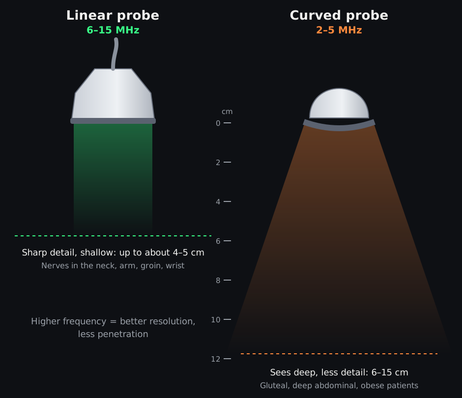 01 · MACHINE, PROBES AND IMAGEPrinciples of ultrasound
How the ultrasound machine makes a picture, which probe to choose, and how to get a clear image of the target.
-
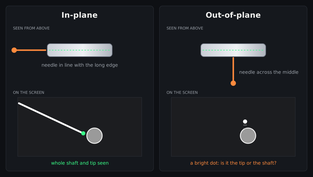 02 · NEEDLES, PLANES AND HAND–EYE SKILLBasics of needling
Choosing the needle, in-plane and out-of-plane approaches, keeping the tip in view, and safe advancement.
-
 03 · PRINCIPLES OF ULTRASOUND-GUIDED BLOCKSBasics in UGRA
The safe routine for every ultrasound-guided regional anaesthesia (UGRA) block, from planning to aftercare.
-
 04 · WHAT TISSUES LOOK LIKEIdentifying structures in UGRA
How muscles, fascia, vessels, bone, pleura and nerves look on ultrasound, and how to be sure of a nerve before you inject.
-
 05 · DRUGS, DOSES AND SAFETYLocal anaesthetics
How local anaesthetics work, which one to choose, safe maximum doses, adjuvants and toxicity.
Based on the pre-course material of the Frimley Ultrasound Guided Plan A Nerve Blocks course (course directors Dr Suresh Jeyaraj, Dr Madan Narayanan and Dr Inna Kuchynska), rewritten and expanded for this website. All diagrams are original. For education: it does not replace supervised training, local guidelines or clinical judgement.