A routine for finding things
- Start with something big and certain: a bone, an artery, a big muscle. Then find the nerve by its relationship to it.
- Optimise depth, gain and focus for the target.
- Slide, tilt and rotate to confirm what you see: a structure that looks like a nerve must behave like one.
- Use light pressure, and colour Doppler whenever a dark circle could be a vessel.
Muscle and fascia
- Muscle: hypoechoic (dark) with bright speckles or streaks (a 'starry night' in short axis, feathery lines in long axis).
- Fascia: bright lines between muscle layers. Many blocks are injections between fascial layers (fascia iliaca, transversus abdominis plane, erector spinae, serratus). Seeing the fascia lift or split as you inject confirms the right plane.

Muscle is grey with fine bright streaks; fascia is a bright line around and between muscles.
Original schematic illustration © Nerve Blocks. Not to scale.
Blood vessels
- Arteries: round, black, pulsatile, hard to compress, often with a bright area behind them (enhancement).
- Veins: black, oval or irregular, close with gentle pressure. Easy to miss if you press too hard.
- Use colour Doppler to check the needle path, not just the target. Small vessels can lie right beside nerves.

Both are black. Arteries pulsate and stay open; veins are oval and close with gentle pressure.
Original schematic illustration © Nerve Blocks. Not to scale.
Bone
- Bright surface line with complete acoustic shadow underneath.
- Useful landmarks: ribs (intercostal, serratus, erector spinae), transverse processes (paravertebral, erector spinae, quadratus lumborum), humerus, femur.
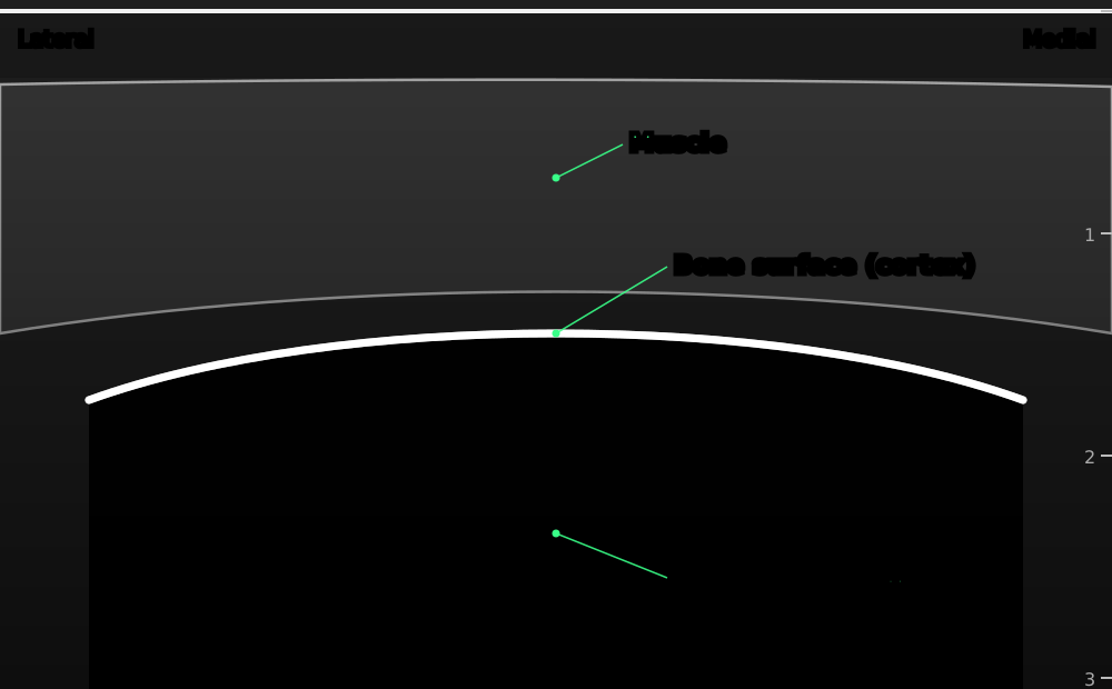
A very bright line with total black shadow underneath: a reliable landmark and a safe 'floor' for many blocks.
Original schematic illustration © Nerve Blocks. Not to scale.
Pleura and lung
- A bright line slightly deeper than the ribs, shimmering as the lung slides with breathing.
- Keep the pleura in view and the needle away from it in chest wall, supraclavicular and paravertebral blocks.
- Lung sliding that disappears after a block suggests a pneumothorax: see our lung ultrasound page.

The pleura is a bright line just deeper than the ribs that slides with each breath.
Original schematic illustration © Nerve Blocks. Not to scale.
Nerves
- Short axis (transverse): round, oval or triangular 'honeycomb': dark fascicles in bright connective tissue. This is the view used for most blocks.
- Long axis: bright parallel lines along the length of the nerve.
- Neck roots (interscalene) look like dark circles with a thin bright rim: little connective tissue.
- Peripheral nerves further out (median, ulnar, radial, femoral, sciatic) look brighter, because they have more connective tissue.
- Examples: brachial plexus between the scalene muscles; median, ulnar and radial nerves around the axillary artery; femoral nerve lateral to the femoral artery under the fascia iliaca; sciatic nerve as a large, bright oval behind the knee or in the buttock.

Short axis: a 'honeycomb' of dark fascicles in bright connective tissue. Long axis: long, parallel lines.
Original schematic illustration © Nerve Blocks. Not to scale.
Is it really a nerve?
- Follow it: slide up and down. A nerve keeps its course and size; a tendon becomes muscle, and a lymph node ends.
- Tilt: a nerve's brightness changes a little with angle, a tendon's a lot.
- Ask the patient to move the fingers or toes: tendons slide, nerves barely move.
- Press gently: veins close, nerves don't.
- Doppler: no flow.
- Check it fits the expected anatomy before you inject, and watch the first 1 mL spread around it.
