Choosing the needle
- Echogenic needles have a textured or coated shaft that reflects more sound, so they are easier to see, especially at steep angles.
- Gauge: 22 G suits most single-shot blocks. 24–25 G for small superficial blocks, 18–20 G Tuohy for catheters.
- Length: 50 mm for superficial blocks (interscalene, axillary, femoral). 80–100 mm for deeper ones (popliteal, erector spinae, gluteal sciatic); 120–150 mm for very deep blocks.
- Short bevel (30–45°): tends to push nerves aside rather than enter them, and gives a better 'feel' of fascia. Insulated needles let you use a nerve stimulator as a safety monitor.
In-plane and out-of-plane
- In-plane: the needle goes in line with the long edge of the probe, so you see the whole shaft and the tip. The best choice for beginners and for most blocks.
- Out-of-plane: the needle crosses the beam in the middle of the probe and shows as a bright dot. The dot may be the shaft, not the tip. Use 'walk-down' (steep then shallower) and small injections of fluid to track the tip. Useful for catheters and superficial targets.
- The steeper the needle, the harder it is to see. Enter the skin a little away from the probe so the needle runs flatter, or heel the probe to bring the beam closer to 90° to the needle.
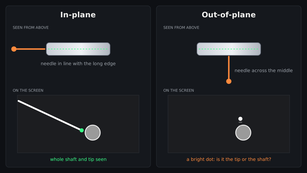
In-plane shows the whole needle; out-of-plane shows only a dot where the needle crosses the beam.
Original diagram © Nerve Blocks.
Hand–eye coordination
- Anchor your probe hand on the patient: little finger or heel of the hand on the skin. The probe must not drift while you move the needle.
- Look at the screen, not your hands, while advancing. Look at your hands only to realign.
- If you lose the tip: stop. Don't advance. Keep the needle still and slide or tilt the probe until the needle comes back, then align again.
- Small in-and-out jiggles of the needle, or a tiny injection of fluid (hydrolocation), confirm where the tip is.
- Move one thing at a time: the probe or the needle, never both.
Advancing safely
- Aim for the tissue plane next to the nerve, not the nerve itself.
- Advance in small steps, only when you can see the tip.
- Aspirate before every injection, then give 0.5–1 mL first to see where the fluid goes, then the rest in 3–5 mL aliquots, aspirating between them.
- Stop if the patient feels sharp pain or paraesthesia, if injection is hard (high pressure), if a nerve swells, or if you can't see the spread. Reposition before you inject again.

Aim the tip tangentially, beside the nerve. A needle aimed straight at a nerve can pierce it.
Original diagram © Nerve Blocks.
Ergonomics
- Put the machine on the far side of the patient, so you look straight past the block site to the screen.
- Raise or lower the bed so your forearms are relaxed and your wrists straight. Sit if you can.
- Probe orientation matches the screen: needle entering from the left of the probe appears from the left of the screen.
- Have the drugs drawn up, labelled and within reach, with an assistant to inject if you prefer.

Set yourself up before you scrub: you, the block site and the screen in one line.
Original diagram © Nerve Blocks.
How to practise
- Practise on a gel phantom or a meat model (for example chicken breast or pork) before patients.
- Do both in-plane and out-of-plane approaches, and practise finding a lost tip.
- Record clips of your blocks and review them with a trainer.
References
- Marhofer P, Chan VWS. Ultrasound-guided regional anesthesia: current concepts and future trends. Anesth Analg 2007;104:1265–9. PubMed