Level of difficulty
Basic. The foramen is easy to feel or see on ultrasound and the injection is shallow.
Anatomy
The infraorbital nerve is the end branch of the maxillary nerve (V2). It runs forwards in the floor of the orbit and comes out on the face through the infraorbital foramen, about 0.5–1 cm below the lower orbital rim, in line with the pupil.
On the face it divides into branches to the lower eyelid, the side of the nose and the upper lip (the inferior palpebral, nasal and superior labial branches).
The foramen lies under levator labii superioris. In infants it sits about halfway between the corner of the mouth and the middle of the lower eyelid.
The infraorbital artery and vein leave the foramen with the nerve.
Surface anatomy
- Lower orbital rim in line with the pupil.
- The foramen is about 0.5–1 cm below the rim.
- Infants: halfway between the corner of the mouth and the middle of the lower eyelid.
Indications
- Cleft lip repair in infants (bilateral for bilateral clefts).
- Upper lip, nose and cheek surgery, including skin lesions and lacerations.
- Endoscopic sinus surgery and septorhinoplasty, as part of the analgesia plan.
- Upper front teeth and gum procedures (intraoral approach).
Nerves blocked
- Infraorbital nerve (V2): inferior palpebral, external nasal and superior labial branches
- Often the anterior superior alveolar nerve (front upper teeth)
Landmark technique
Surface anatomy, relevant structures and landmarks
Feel the foramen below the orbital rim in line with the pupil.
Positioning
Supine. Keep a finger on the orbital rim to protect the eye.
Block technique
- Extraoral: insert the needle just below the foramen, aiming upwards towards it, until bone is touched near the opening. Do not enter the canal. Aspirate and inject.
- Intraoral (used often in children): lift the upper lip, insert the needle into the mucosa above the canine or first premolar and advance upwards, parallel to the bone, towards the finger on the foramen.
- Press on the site afterwards to reduce swelling.
Needle choice
25–27 G, 25 mm.
Neurostimulation technique
Not used.
Choice of local anaesthetic (including volume)
- Infants: 0.5–1 mL of 0.25% bupivacaine or levobupivacaine each side (keep within weight-based limits).
- Adults: 1–3 mL of 1–2% lidocaine with adrenaline, or 0.25–0.5% bupivacaine or levobupivacaine.
Ultrasound technique
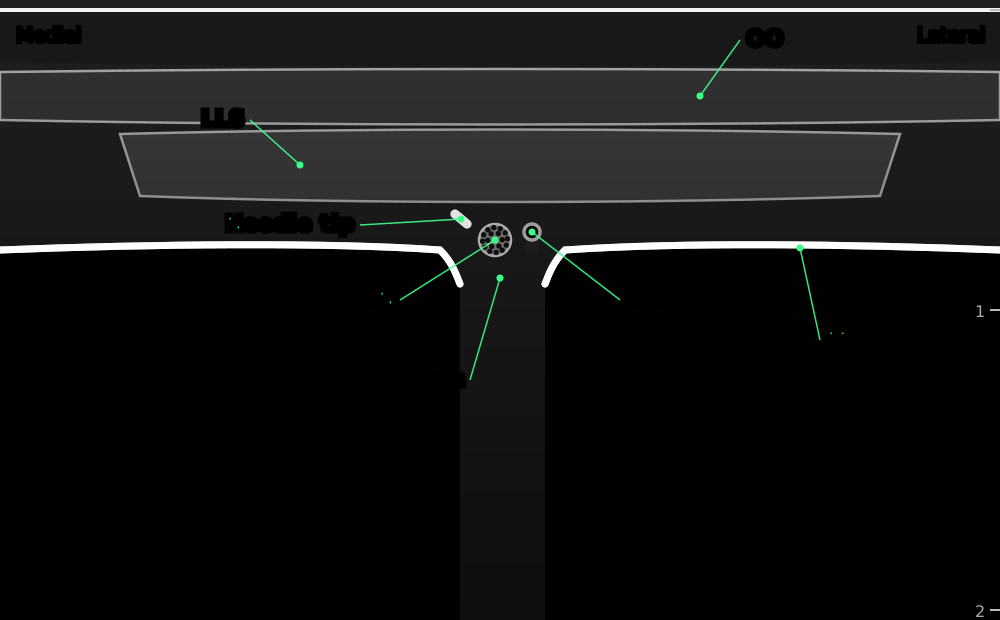

Transverse view just below the lower orbital rim. The bright line of the maxilla has a gap at the infraorbital foramen, where the nerve and artery come out under levator labii superioris.
- OO
- orbicularis oculi
- LLS
- levator labii superioris, lying over the foramen
- Maxilla
- front of the maxilla (bright line with shadow)
- Foramen
- infraorbital foramen: a gap in the bone line
- ION
- infraorbital nerve
- IOA
- infraorbital artery (colour Doppler)
- Needle tip
- needle seen end-on (out of plane), coming from below
Positioning
Supine with the operator at the head end.
Scanning technique
- Place a small linear probe horizontally just below the lower orbital rim.
- Slide down slowly until the smooth bright line of the maxilla shows a break: the foramen.
- Colour Doppler shows the infraorbital artery.
Structures to identify
- Maxilla (bright line)
- Infraorbital foramen
- Infraorbital nerve and artery
- Levator labii superioris and orbicularis oculi
Block performance
Position / ergonomics
Stand at the head of the bed.
Probe
Small linear high-frequency (hockey-stick) probe.
Settings
Superficial preset, depth 1.5–2 cm.
Depth
0.3–1 cm.
Needle size
25–27 G, 25 mm.
Needling technique
Out of plane from below, aiming at the foramen.
Optimal needle-tip position
At the opening of the foramen, outside the canal.
Drug choice
See above.
Volume
0.5–1 mL in infants; 1–3 mL in adults.
Optimal current if nerve stimulation is used
Not used.
Muscle responses acceptable
Not applicable.
Testing block success
- Numb upper lip, side of the nose and lower eyelid
Continuous techniques
Not used.
Contraindications to the block
Absolute
- Patient refusal
- Infection at the injection site (skin or mouth)
- Allergy to the drugs used
Relative
- Fracture of the orbital floor or maxilla at the site
- Coagulopathy (usually acceptable: superficial and compressible)
Side effects and complications
Side effects
- Numb upper lip (warn about biting it)
- Cheek swelling and bruising
Complications
- Injection into the orbit (needle passed up the canal)
- Intravascular injection
- Nerve injury from injecting into the canal (rare)
Pearls of wisdom
- Keep a finger on the orbital rim and never thread the needle up the canal.
- In infants, a small volume is enough: 0.5–1 mL each side.
- For cleft palate surgery the infraorbital block alone is not enough: consider a suprazygomatic maxillary block.
- Warn patients and parents about the numb lip after surgery.
Review of literature
- Feriani and colleagues (Cochrane, 2016) reviewed infraorbital nerve block for cleft lip repair in children. Small trials suggested better pain relief and less need for other analgesics, but the evidence was of low quality.
- Echaniz and colleagues (2019) found bilateral suprazygomatic maxillary blocks gave better analgesia than infraorbital plus palatine blocks for combined cleft lip and palate repair.
- Allam and colleagues (2018) described the ultrasound anatomy of the infraorbital foramen.
Comparison with other blocks
| Block | Area numbed | Depth | Main use |
|---|---|---|---|
| Infraorbital | Lower eyelid, cheek, side of nose, upper lip | Shallow | Cleft lip, lip and nose surgery |
| Maxillary (suprazygomatic) | Whole V2 area including palate | Deep | Cleft palate |
| Mental | Lower lip and chin | Shallow | Lower lip and chin surgery |
References
- Feriani G, Hatanaka E, Torloni MR, da Silva EM. Infraorbital nerve block for postoperative pain following cleft lip repair in children. Cochrane Database Syst Rev 2016;4:CD011131. PubMed
- Echaniz G, De Miguel M, Merritt G, et al. Bilateral suprazygomatic maxillary nerve blocks vs. infraorbital and palatine nerve blocks in cleft lip and palate repair: a double-blind, randomised study. Eur J Anaesthesiol 2019;36:40–47. PubMed
- Allam AE, Khalil AAF, Eltawab BA, Wu WT, Chang KV. Ultrasound-guided intervention for treatment of trigeminal neuralgia: an updated review of anatomy and techniques. Pain Res Manag 2018;2018:5480728. PubMed
Test yourself: Infraorbital nerve block
4 questions on this page and up to 4 on the scan: name the structure at the numbered dot. Score 70% or more to add this topic to My learning and your certificate. You can try again as often as you like.
New to ultrasound? Start with the Basics of ultrasound or try the scan explorer. Compare this block with another.