1. Home
  2. Ultrasound in anaesthesia
  3. Scan explorer
INTERACTIVE · ULTRASOUND IN ANAESTHESIA

Scan explorer

Move the probe along a nerve's journey and watch the ultrasound picture change, stop by stop.

Slide the probe along a nerve's journey and watch the ultrasound picture change. Drag the probe, press a numbered stop, or use the slider and the arrow keys. Each stop shows the real ultrasound scan (Anaesthesia Sonoanatomy (ANSO) app), our own labelled schematic illustration, or both side by side: choose with the buttons above the picture.

Brachial plexus: neck to axilla

Follow the plexus from the roots in the neck to the terminal nerves in the axilla.

CHEST ARM ABDUCTED, PALM UP CLAVICLE
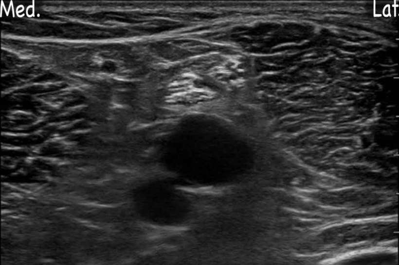
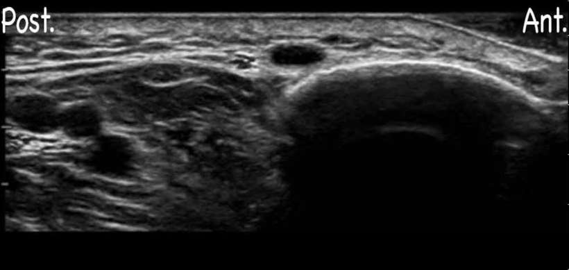
Ultrasound scan: InterscaleneSuperior trunk block: labelled schematic ultrasound illustrationUltrasound scan: SupraclavicularUltrasound scan: CostoclavicularUltrasound scan: InfraclavicularAxillary brachial plexus block: labelled schematic ultrasound illustrationSCAN
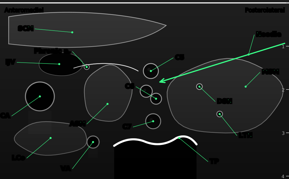
Interscalene brachial plexus block: labelled schematic ultrasound illustrationSuperior trunk block: labelled schematic ultrasound illustrationSupraclavicular brachial plexus block: labelled schematic ultrasound illustrationCostoclavicular brachial plexus block: labelled schematic ultrasound illustrationInfraclavicular brachial plexus block: labelled schematic ultrasound illustrationAxillary brachial plexus block: labelled schematic ultrasound illustrationILLUSTRATION
1 Interscalene

C5, C6 and C7 roots lie stacked between scalenus anterior and medius.

Open the Interscalene page ›
Illustrations: original schematic illustrations © Nerve Blocks. Not to scale.
Ultrasound images: Anaesthesia Sonoanatomy (ANSO) app.

Median nerve: axilla to wrist

Trace the median nerve down the arm, from beside the axillary artery to the carpal tunnel.

CHEST ARM ABDUCTED, PALM UP CLAVICLE
Axillary brachial plexus block: labelled schematic ultrasound illustrationUltrasound scan: Mid-humeralUltrasound scan: ElbowUltrasound scan: ForearmWrist block, median nerve: labelled schematic ultrasound illustrationSCAN
Axillary brachial plexus block: labelled schematic ultrasound illustrationMid-humeral block, medial view: labelled schematic ultrasound illustrationElbow block, median nerve: labelled schematic ultrasound illustrationMid-forearm block, median nerve: labelled schematic ultrasound illustrationWrist block, median nerve: labelled schematic ultrasound illustrationILLUSTRATION
1 Axillary

The median nerve starts next to the axillary artery.

Open the Axillary page ›
Illustrations: original schematic illustrations © Nerve Blocks. Not to scale.
Ultrasound images: Anaesthesia Sonoanatomy (ANSO) app.

Sciatic nerve: buttock to ankle

Follow the sciatic nerve from the pelvis to the knee, then its tibial branch to the ankle.

PRONE: BACK OF THE LEG KNEE ANKLE
Ultrasound scan: Parasacral sciaticUltrasound scan: Subgluteal sciaticUltrasound scan: Popliteal sciaticUltrasound scan: AnkleSCAN
Parasacral sciatic block: labelled schematic ultrasound illustrationSubgluteal sciatic block: labelled schematic ultrasound illustrationPopliteal sciatic block at the bifurcation: labelled schematic ultrasound illustrationAnkle block, tibial nerve: labelled schematic ultrasound illustrationILLUSTRATION
1 Parasacral sciatic

The sacral plexus leaves the pelvis through the greater sciatic notch, deep to gluteus maximus.

Open the Parasacral sciatic page ›
Illustrations: original schematic illustrations © Nerve Blocks. Not to scale.
Ultrasound images: Anaesthesia Sonoanatomy (ANSO) app.

Femoral and saphenous nerves: groin to ankle

Follow the femoral nerve at the groin and its saphenous branch down to the ankle.

SUPINE: FRONT OF THE LEG KNEE ANKLE
Ultrasound scan: FemoralUltrasound scan: Femoral triangleUltrasound scan: Adductor canalUltrasound scan: AnkleSCAN
Femoral nerve block: labelled schematic ultrasound illustrationFemoral triangle block (distal): labelled schematic ultrasound illustrationAdductor canal block: labelled schematic ultrasound illustrationAnkle block, saphenous nerve: labelled schematic ultrasound illustrationILLUSTRATION
1 Femoral

At the groin the femoral nerve lies lateral to the femoral artery, under fascia iliaca.

Open the Femoral page ›
Illustrations: original schematic illustrations © Nerve Blocks. Not to scale.
Ultrasound images: Anaesthesia Sonoanatomy (ANSO) app.

Share the scan explorer with your trainees.

Share

For learning anatomy only. Real scans vary between patients; always scan and confirm before you inject.