Level of difficulty
Intermediate. The target plane is easy to see, but the carotid artery, internal jugular vein and the prevertebral fascia are close, so careful needle control is needed.
Anatomy
The neck has three layers of deep cervical fascia that matter here: the investing layer, which wraps sternocleidomastoid; the prevertebral layer, which covers the scalene and levator scapulae muscles and the roots of the plexuses; and the carotid sheath.
The sensory branches of the cervical plexus leave the plexus under the prevertebral fascia, pass through it, and then run in the space between it and the investing fascia (the posterior cervical space) before winding round the back edge of sternocleidomastoid.
An intermediate block puts the drug in this space, under sternocleidomastoid but on top of the prevertebral fascia. The prevertebral fascia should keep the drug away from the phrenic nerve and the roots, which lie deep to it.
Drug in this space often spreads medially towards the carotid sheath, which may help during carotid surgery.
Surface anatomy
- Mastoid process.
- Back edge of sternocleidomastoid at its midpoint (about the level of the thyroid cartilage, C4).
- External jugular vein.
- Carotid pulse (medial, to stay away from).
Indications
- Carotid endarterectomy under regional anaesthesia (with surgical top-up near the jaw and the carotid bifurcation).
- Thyroid and parathyroid surgery, for analgesia.
- Neck dissection skin and lymph node surgery.
- Clavicle surgery, together with an interscalene or superior trunk block.
Nerves blocked
- Lesser occipital, great auricular, transverse cervical and supraclavicular nerves (C2–C4)
- Sometimes branches of the ansa cervicalis and small twigs to the carotid sheath
Landmark technique
Surface anatomy, relevant structures and landmarks
Mark the midpoint of the back edge of sternocleidomastoid, as for the superficial block.
Positioning
Supine with the head turned 30–45° away.
Block technique
- Insert a short-bevel needle at the back edge of the muscle, aiming under it.
- Feel a 'pop' as the needle passes through the investing fascia, then inject 10–15 mL after aspiration.
- Ultrasound is strongly preferred, because the depth of the plane varies.
Needle choice
22 G short-bevel, 50 mm.
Neurostimulation technique
Not used.
Choice of local anaesthetic (including volume)
- 10–15 mL of 0.25–0.5% levobupivacaine, 0.375–0.5% ropivacaine or a lidocaine and long-acting mixture for carotid surgery.
- Keep the total dose (including the surgeon's top-up) within the maximum: use the dose calculator.
Ultrasound technique
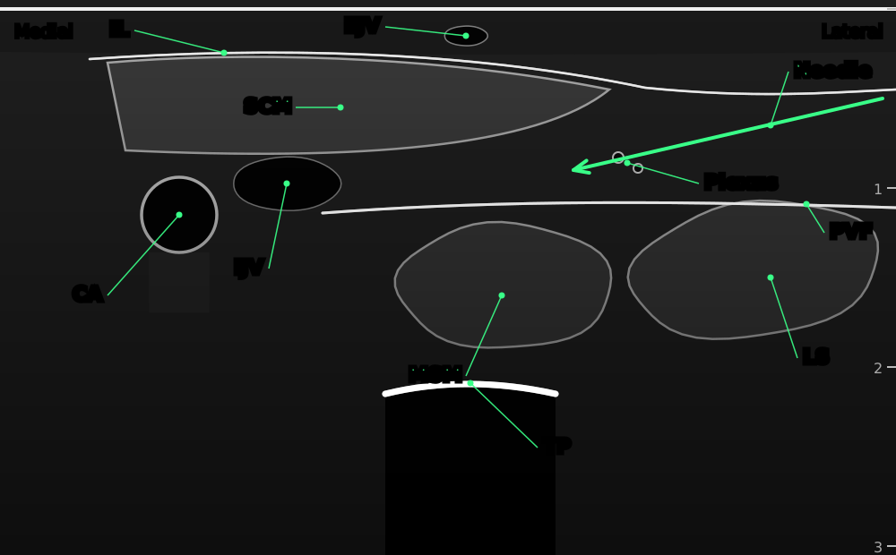

Transverse view at C4. The needle passes under the back edge of sternocleidomastoid and local anaesthetic spreads between the muscle and the prevertebral fascia, separating the two.
- SCM
- sternocleidomastoid
- IL
- investing layer of the deep cervical fascia: the white layer over sternocleidomastoid
- EJV
- external jugular vein
- Plexus
- branches of the cervical plexus emerging at the posterior border of SCM
- CA
- common carotid artery
- IJV
- internal jugular vein
- PVF
- prevertebral fascia: the needle stays superficial to it
- MSM
- middle scalene muscle
- LS
- levator scapulae
- TP
- C4 transverse process with acoustic shadow
- Needle
- in plane from lateral, under the posterior edge of SCM
Positioning
As above, with the screen on the other side of the patient.
Scanning technique
- Place the probe across the neck at C4 over sternocleidomastoid; find the carotid artery and internal jugular vein under the muscle.
- Slide laterally until the tapering back edge of sternocleidomastoid is in the middle of the screen.
- Identify the bright prevertebral fascia over levator scapulae and the scalene muscles, and the small nerve branches lying on it.
- Use colour Doppler to check the needle path.
Structures to identify
- Sternocleidomastoid and its investing fascia
- Prevertebral fascia
- Cervical plexus branches in the posterior cervical space
- Levator scapulae and middle scalene
- Carotid artery and internal jugular vein (medial)
- External jugular vein (superficial)
Block performance

Position / ergonomics
Stand at the head of the bed or on the side of the block.
Probe
Linear, high frequency.
Settings
Nerve or small parts preset, depth 3 cm.
Depth
1–2.5 cm.
Needle size
22 G, 50 mm short-bevel.
Needling technique
In-plane from lateral to medial, passing under the back edge of the muscle.
Optimal needle-tip position
Under sternocleidomastoid and on top of the prevertebral fascia. The drug should lift the muscle off the fascia. If it spreads under the prevertebral fascia, stop and pull back: that is a deep block, with a higher risk of phrenic nerve block.
Drug choice
As above.
Volume
10–15 mL.
Optimal current if nerve stimulation is used
Not used.
Muscle responses acceptable
Not applicable.
Testing block success
- Loss of sensation over the front and side of the neck, ear lobe and clavicle
- Comfort when the surgeon cuts the skin
Continuous techniques
Rarely used. Catheters in this space have been described for major neck surgery but are not standard.
Contraindications to the block
Absolute
- Patient refusal
- Infection at the injection site
- Allergy to the drugs used
Relative
- Coagulopathy or anticoagulant timing outside guideline limits (the injection is deeper and near large vessels)
- Contralateral phrenic nerve or recurrent laryngeal nerve palsy
- Severe respiratory disease, for bilateral blocks
- Distorted neck anatomy (previous surgery or radiotherapy)
Side effects and complications
Side effects
- Numb neck and ear
- Hoarse voice or difficulty swallowing if the drug spreads to the vagus or recurrent laryngeal nerve
- Horner's syndrome
Complications
- Phrenic nerve block (less than with a deep block, but possible)
- Carotid or jugular puncture and haematoma
- Intravascular injection and local anaesthetic toxicity
- Spread to the brachial plexus
Pearls of wisdom
- Watch the drug lift sternocleidomastoid off the prevertebral fascia; if it disappears deep, stop.
- For carotid surgery, plan the surgeon's top-up into the total dose from the start.
- Avoid bilateral intermediate blocks in patients who rely on both hemidiaphragms.
- Trials comparing it with a subcutaneous (superficial) block for carotid surgery found similar results, so choose the one you do most safely.
Review of literature
- Pandit and colleagues (2007) found superficial and intermediate blocks safer than deep or combined blocks for carotid endarterectomy, with fewer conversions to general anaesthesia.
- Ramachandran and colleagues (2011) randomised 44 patients having carotid endarterectomy to an intermediate or a subcutaneous block: the surgeon's lidocaine top-up and patient satisfaction were similar.
- Kim and colleagues (2018) explained why the name 'intermediate' has caused confusion, and described the fascial compartments seen on ultrasound.
Comparison with other blocks
| Block | Injection site | Phrenic palsy risk | Main use |
|---|---|---|---|
| Superficial cervical plexus | Under platysma | Very low | Thyroid, superficial neck |
| Intermediate cervical plexus | Between SCM and prevertebral fascia | Low | Carotid endarterectomy |
| Deep cervical plexus | C2–C4 transverse processes | High | Rarely used now |
| Interscalene | Between anterior and middle scalene | Very high | Shoulder |
References
- Pandit JJ, Satya-Krishna R, Gration P. Superficial or deep cervical plexus block for carotid endarterectomy: a systematic review of complications. Br J Anaesth 2007;99:159–169. PubMed
- Ramachandran SK, Picton P, Shanks A, Dorje P, Pandit JJ. Comparison of intermediate vs subcutaneous cervical plexus block for carotid endarterectomy. Br J Anaesth 2011;107:157–163. PubMed
- Kim JS, Ko JS, Bang S, Kim H, Lee SY. Cervical plexus block. Korean J Anesthesiol 2018;71:274–288. PubMed
Test yourself: Intermediate cervical plexus block
4 questions on this page and up to 4 on the scan: name the structure at the numbered dot. Score 70% or more to add this topic to My learning and your certificate. You can try again as often as you like.
New to ultrasound? Start with the Basics of ultrasound or try the scan explorer. Compare this block with another.