Level of difficulty
Advanced. The fossa is deep, small and full of blood vessels, and the needle passes close to the orbit. The suprazygomatic approach in children is well described and has a good safety record in experienced hands.
Anatomy
The maxillary nerve (V2) is the second division of the trigeminal nerve. It leaves the skull through the foramen rotundum into the pterygopalatine fossa, a small space behind the maxilla, in front of the pterygoid plates and below the greater wing of the sphenoid.
In the fossa it gives branches to the pterygopalatine ganglion (palatine and nasal nerves), the zygomatic nerve and the posterior superior alveolar nerves, then continues forwards as the infraorbital nerve.
The fossa also contains the third part of the maxillary artery and its branches, which can be seen with colour Doppler.
The suprazygomatic approach enters at the angle between the upper edge of the zygomatic arch and the back edge of the frontal process of the zygoma, passes through temporalis and over the greater wing of the sphenoid into the fossa. The infrazygomatic approach passes below the arch, through the coronoid notch of the mandible and the lateral pterygoid muscle.
Surface anatomy
- Zygomatic arch.
- Frontozygomatic angle (where the arch meets the side of the orbit).
- Coronoid notch of the mandible (below the arch; open the mouth slightly to feel it).
- Lateral edge of the orbit.
Indications
- Cleft palate repair in infants (bilateral suprazygomatic block).
- Maxillary and upper jaw surgery, including orthognathic surgery and fractures.
- Upper dental and sinus surgery.
- Facial pain clinics: trigeminal neuralgia in the V2 area, sphenopalatine ganglion block for headache.
Nerves blocked
- Maxillary nerve (V2) and its branches: greater and lesser palatine, nasopalatine, posterior superior alveolar, zygomatic and infraorbital nerves
- Pterygopalatine (sphenopalatine) ganglion
Landmark technique
Surface anatomy, relevant structures and landmarks
Suprazygomatic: the entry point is at the angle formed by the upper edge of the zygomatic arch and the back edge of the frontal process of the zygoma. Infrazygomatic: the entry point is in the coronoid notch, just below the middle of the arch.
Positioning
Supine with the head neutral (children are usually asleep).
Block technique
- Suprazygomatic: insert the needle at right angles to the skin until it touches the greater wing of the sphenoid (about 10–15 mm in infants), then turn it towards the philtrum and slightly down and advance a further 10–15 mm into the fossa.
- Infrazygomatic: insert the needle through the coronoid notch, aiming slightly forwards and up towards the fossa, to a depth of about 4–5 cm in adults.
- Aspirate carefully and inject slowly in small amounts.
Needle choice
22–25 G short-bevel, 35 mm in children and 50 mm in adults.
Neurostimulation technique
Not usually used (sensory nerve).
Choice of local anaesthetic (including volume)
- Children: 0.15 mL/kg of 0.2% ropivacaine (or 0.25% levobupivacaine) each side.
- Adults: 3–5 mL of 0.5% ropivacaine or 0.25–0.5% levobupivacaine each side.
- Check the total dose for bilateral blocks: use the dose calculator.
Ultrasound technique
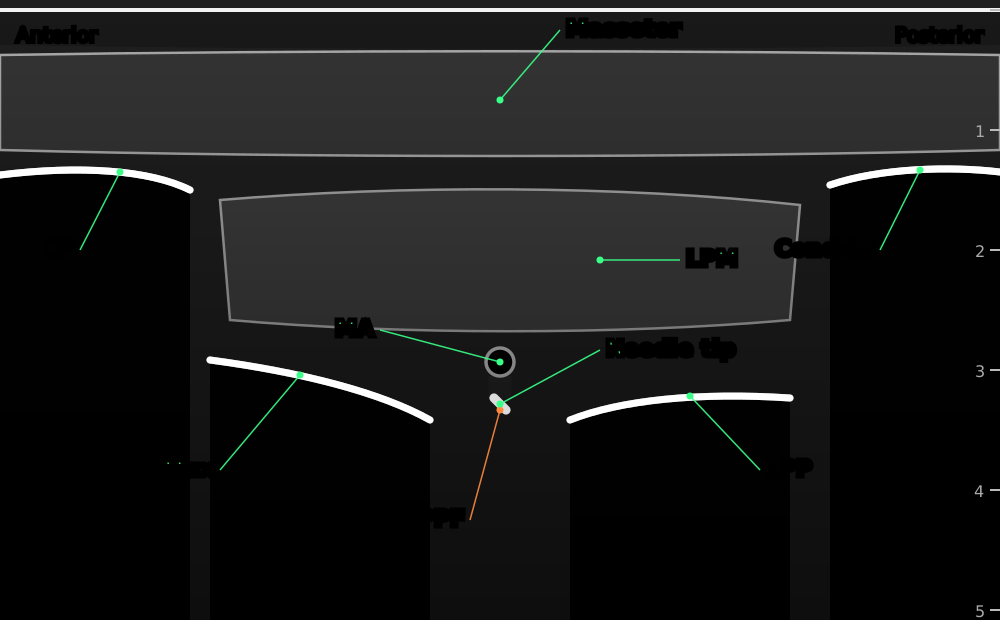
Probe under the zygomatic arch, over the maxilla. The needle comes from above the arch and is seen as a bright dot as it enters the fossa.
- Masseter
- masseter muscle
- CP
- coronoid process of the mandible
- Condyle
- condyle of the mandible
- LPM
- lateral pterygoid muscle
- Max
- back wall of the maxilla
- LPP
- lateral pterygoid plate
- PPF
- pterygopalatine fossa, filled with local anaesthetic
- MA
- maxillary artery: check with colour Doppler
- Needle tip
- needle seen end-on (out of plane), coming from above the zygomatic arch


Same window under the arch. The needle passes in plane through the notch of the mandible and the lateral pterygoid muscle into the fossa.
- Masseter
- masseter muscle
- CP
- coronoid process of the mandible
- Condyle
- condyle of the mandible
- LPM
- lateral pterygoid muscle
- Max
- back wall of the maxilla
- LPP
- lateral pterygoid plate
- PPF
- pterygopalatine fossa, filled with local anaesthetic
- MA
- maxillary artery: check with colour Doppler
- Needle
- in plane from behind, through the mandibular notch and the lateral pterygoid muscle
Positioning
Supine with the head turned slightly away. In infants, a hockey-stick probe fits under the zygoma.
Scanning technique
- Place the probe below the zygomatic arch, over the maxilla, tilted slightly upwards (the infrazygomatic window).
- Find the bright curved line of the back of the maxilla and, behind it, the lateral pterygoid plate. The gap between them is the pterygopalatine fossa.
- The lateral pterygoid muscle lies over the fossa. Use colour Doppler to see the maxillary artery.
- Suprazygomatic approach: keep the probe here and bring the needle in out of plane from above the arch; watch the drug fill the fossa.
- Infrazygomatic approach: bring the needle in plane from the back (or front) edge of the probe through the lateral pterygoid muscle.
Structures to identify
- Zygomatic arch
- Maxilla (back wall)
- Lateral pterygoid plate
- Lateral pterygoid muscle
- Pterygopalatine fossa
- Maxillary artery
- Coronoid process of the mandible
Block performance
Position / ergonomics
Stand at the head of the bed, with the screen on the opposite side.
Probe
Linear high-frequency, or a hockey-stick probe in infants.
Settings
Small parts preset, depth 4–5 cm, colour Doppler on.
Depth
About 2 cm in infants; 4–4.5 cm in adults.
Needle size
22–25 G, 35–50 mm short-bevel.
Needling technique
Suprazygomatic: out of plane from above the arch. Infrazygomatic: in plane under the arch through the coronoid notch.
Optimal needle-tip position
In the pterygopalatine fossa, away from the maxillary artery. Spread should be seen filling the fossa.
Drug choice
See the landmark section.
Volume
0.15 mL/kg each side in children; 3–5 mL in adults.
Optimal current if nerve stimulation is used
Not used.
Muscle responses acceptable
Not applicable. A jaw (masseter) twitch means the needle is near the mandibular nerve, too far back.
Testing block success
- Numb cheek, upper lip and side of the nose (in awake adults)
- No heart rate response to the surgical incision on the palate
Continuous techniques
Not usually used. Catheters have been described in adults for maxillofacial surgery and pain, but are not routine.
Contraindications to the block
Absolute
- Patient refusal
- Infection at the injection site
- Allergy to the drugs used
Relative
- Coagulopathy (the pterygopalatine fossa is vascular and cannot be compressed)
- Distorted anatomy (facial fractures, tumours)
- Previous surgery in the fossa
Side effects and complications
Side effects
- Numb cheek, upper lip and teeth
- Facial bruising or swelling
Complications
- Puncture of the maxillary artery: haematoma, or pseudoaneurysm later
- Intravascular injection and local anaesthetic toxicity (the head and neck are very vascular)
- Spread into the orbit: double vision or temporary visual disturbance
- Block of the mandibular nerve (needle too far back)
- Infection (rare)
Pearls of wisdom
- In infants, touching the greater wing of the sphenoid before redirecting gives a reliable depth guide.
- Use colour Doppler every time: the maxillary artery runs through the fossa.
- Inject slowly in small amounts and watch the ECG: small volumes into an artery here can reach the brain.
- Bilateral suprazygomatic blocks cut opioid needs after cleft palate repair, which may lower the risk of airway problems.
Review of literature
- Chiono and colleagues (2014) randomised 60 infants having cleft palate repair: bilateral suprazygomatic blocks with 0.15 mL/kg 0.2% ropivacaine halved morphine use over 48 hours compared with saline.
- Sola and colleagues (2012) showed that ultrasound let them see the fossa and the spread of drug in 94% of 50 suprazygomatic blocks in children, with no complications.
- Echaniz and colleagues (2019) found bilateral suprazygomatic blocks gave better analgesia than infraorbital plus palatine blocks for cleft lip and palate repair.
- Anugerah and colleagues (2020) reviewed the ultrasound-guided approaches to the pterygopalatine fossa, and Jerman and colleagues (2023) found the suprazygomatic path passed further from the maxillary artery than the infrazygomatic path in a virtual model.
Comparison with other blocks
| Block | Area numbed | Depth | Main use |
|---|---|---|---|
| Maxillary (suprazygomatic) | Whole V2 area, including palate | Deep | Cleft palate, maxillary surgery |
| Maxillary (infrazygomatic) | Whole V2 area, including palate | Deep | Adult maxillofacial surgery, pain |
| Infraorbital | Lower eyelid, cheek, side of nose, upper lip | Shallow | Cleft lip, nasal and lip surgery |
| Greater palatine (intraoral) | Hard palate | Shallow | Palate surgery (by the surgeon) |
References
- Chiono J, Raux O, Bringuier S, et al. Bilateral suprazygomatic maxillary nerve block for cleft palate repair in children: a prospective, randomized, double-blind study versus placebo. Anesthesiology 2014;120:1362–1369. PubMed
- Sola C, Raux O, Savath L, Macq C, Capdevila X, Dadure C. Ultrasound guidance characteristics and efficiency of suprazygomatic maxillary nerve blocks in infants: a descriptive prospective study. Paediatr Anaesth 2012;22:841–846. PubMed
- Echaniz G, De Miguel M, Merritt G, et al. Bilateral suprazygomatic maxillary nerve blocks vs. infraorbital and palatine nerve blocks in cleft lip and palate repair: a double-blind, randomised study. Eur J Anaesthesiol 2019;36:40–47. PubMed
- Anugerah A, Nguyen K, Nader A. Technical considerations for approaches to the ultrasound-guided maxillary nerve block via the pterygopalatine fossa: a literature review. Reg Anesth Pain Med 2020;45:301–305. PubMed
- Jerman A, Umek N, Cvetko E, et al. Comparison of the feasibility and safety of infrazygomatic and suprazygomatic approaches to pterygopalatine fossa using virtual reality. Reg Anesth Pain Med 2023;48:359–364. PubMed
Test yourself: Maxillary nerve block: suprazygomatic and infrazygomatic approaches
4 questions on this page and up to 4 on the scan: name the structure at the numbered dot. Score 70% or more to add this topic to My learning and your certificate. You can try again as often as you like.
New to ultrasound? Start with the Basics of ultrasound or try the scan explorer. Compare this block with another.