Level of difficulty
Basic. The foramen is easy to find by feel or ultrasound, and the injection is shallow.
Anatomy
The mental nerve is the end branch of the inferior alveolar nerve, from the mandibular division of the trigeminal nerve (V3).
It leaves the mandible through the mental foramen, which in adults lies below the second premolar (or between the two premolars), about halfway between the gum line and the lower border of the jaw, and roughly in line with the pupil.
The foramen opens upwards and backwards. The nerve divides into branches to the skin of the chin and to the skin and lining of the lower lip.
In children the foramen is lower on the jaw, and in people without teeth it moves up towards the top of the bone.
Surface anatomy
- Second lower premolar tooth.
- Midway between the gum line and the lower border of the mandible.
- In line with the pupil and the supraorbital and infraorbital foramina.
Indications
- Lower lip and chin surgery: lacerations, skin lesions, lip biopsy.
- Chin augmentation and other cosmetic procedures.
- Lower front teeth and gum procedures (incisive branch, intraoral approach).
Nerves blocked
- Mental nerve (V3)
- Often the incisive nerve (lower canine and incisors)
Landmark technique
Surface anatomy, relevant structures and landmarks
Feel the foramen below the second premolar, halfway down the jaw.
Positioning
Supine or sitting.
Block technique
- Intraoral: pull the lower lip down, insert the needle into the fold between lip and gum next to the second premolar and advance 5–10 mm towards the foramen. Aspirate and inject 1–2 mL.
- Extraoral: insert the needle through the skin over the foramen, aiming slightly down and forwards to meet the opening. Do not enter the canal.
- Press on the site for a minute.
Needle choice
25–27 G, 25 mm.
Neurostimulation technique
Not used.
Choice of local anaesthetic (including volume)
- 1–2 mL of 1–2% lidocaine with adrenaline, or 0.25–0.5% bupivacaine or levobupivacaine for longer effect.
Ultrasound technique
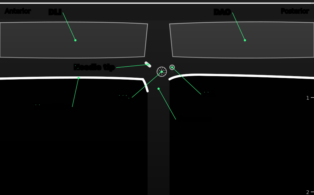

Long-axis view along the body of the mandible below the premolars. The bright line of bone has a gap at the mental foramen, where the mental nerve and vessels come out under the depressor muscles of the lower lip.
- DLI
- depressor labii inferioris
- DAO
- depressor anguli oris
- Mandible
- body of the mandible below the premolars (bright line with shadow)
- Foramen
- mental foramen: a gap in the bone line, opening up and back
- MN
- mental nerve
- MA
- mental artery (colour Doppler)
- Needle tip
- needle seen end-on (out of plane)
Positioning
Supine with the head turned slightly away.
Scanning technique
- Place a small linear probe along the lower jaw, below the premolar teeth.
- Slide along the bone until the bright line breaks: the mental foramen.
- Colour Doppler shows the mental artery.
Structures to identify
- Mandible (bright line)
- Mental foramen
- Mental nerve and artery
- Depressor anguli oris and depressor labii inferioris
Block performance
Position / ergonomics
Stand on the side of the block or at the head of the bed.
Probe
Small linear high-frequency (hockey-stick) probe.
Settings
Superficial preset, depth 1.5–2 cm.
Depth
0.3–1 cm.
Needle size
25–27 G, 25 mm.
Needling technique
Out of plane, aiming at the foramen.
Optimal needle-tip position
At the opening of the foramen, outside the canal.
Drug choice
See above.
Volume
1–2 mL.
Optimal current if nerve stimulation is used
Not used.
Muscle responses acceptable
Not applicable.
Testing block success
- Numb lower lip and chin on that side
Continuous techniques
Not used.
Contraindications to the block
Absolute
- Patient refusal
- Infection at the injection site (skin or mouth)
- Allergy to the drugs used
Relative
- Fracture of the mandible at the foramen
- Coagulopathy (usually acceptable: superficial and compressible)
Side effects and complications
Side effects
- Numb lower lip (warn about biting it or spilling drinks)
- Bruising
Complications
- Intravascular injection
- Nerve injury from injecting into the canal (rare)
- Haematoma
Pearls of wisdom
- The intraoral route is less painful and leaves no skin mark.
- For midline lip surgery, block both sides: fibres cross the midline.
- In people without teeth the foramen moves up towards the top of the jaw.
Review of literature
- Allam and colleagues (2018) reviewed the anatomy of the mental foramen and the ultrasound-guided technique, as part of a review of trigeminal branch blocks.
- Evidence for the mental block comes mainly from dental and dermatological practice; there are few trials in anaesthesia.
Comparison with other blocks
| Block | Area numbed | Division |
|---|---|---|
| Mental | Lower lip and chin | V3 |
| Inferior alveolar (dental) | Lower teeth, lower lip and chin on one side | V3 |
| Infraorbital | Upper lip, side of nose, lower eyelid | V2 |
References
- Allam AE, Khalil AAF, Eltawab BA, Wu WT, Chang KV. Ultrasound-guided intervention for treatment of trigeminal neuralgia: an updated review of anatomy and techniques. Pain Res Manag 2018;2018:5480728. PubMed
Test yourself: Mental nerve block
4 questions on this page and up to 4 on the scan: name the structure at the numbered dot. Score 70% or more to add this topic to My learning and your certificate. You can try again as often as you like.
New to ultrasound? Start with the Basics of ultrasound or try the scan explorer. Compare this block with another.