Level of difficulty
Intermediate. The bony landmarks are easy to see, but the sacrum is a curved, uneven surface and the target plane lies right on the bone. The name and the best injection point are still being worked out.
Anatomy
The back of the sacrum has three bony ridges on each side of the midline: the median sacral crest (fused spinous processes), the intermediate crest (fused articular processes) and the lateral crest. The dorsal sacral foramina lie just outside the intermediate crest.
The multifidus muscle fills the groove beside the median crest, covered by the tendinous aponeurosis of erector spinae. Local anaesthetic placed between these muscles and the bone is in the same plane as the lumbar and thoracic erector spinae plane.
From here it reaches the dorsal rami of the sacral nerves, which supply the skin over the sacrum and buttock. Spread through the dorsal foramina may also reach the ventral rami and the sacral epidural space. This probably explains the perineal and genital analgesia, but the exact mechanism is still debated.
Contrast studies show spread up to the lumbar levels, which explains reports of analgesia for hip and lumbar spine surgery.
Because the injection is deep to multifidus, some authors call it the sacral multifidus plane block. The target is the same.

Left: probe placed over the sacrum. Right: the back of the sacrum. Multifidus and erector spinae lie over the median and intermediate crests, and the dorsal sacral nerves leave through the dorsal foramina beside them.

The sacral plexus seen from inside the pelvis (front, ventral rami) and the dorsal sacral rami S1–S4 behind the sacrum. The pudendal nerve (S2–S4) supplies the perineum.
Surface anatomy
- Posterior superior iliac spines (PSIS): a line between them crosses S2.
- Median sacral crest in the midline.
- Sacral hiatus and cornua at the lower end (for orientation).
- Natal cleft.
Indications
- Anorectal surgery: haemorrhoidectomy, fistula, fissure and pilonidal sinus surgery.
- Children's genital and urological surgery: circumcision and hypospadias repair, as an alternative to a caudal block.
- Transurethral and other perineal surgery.
- Lumbar spine surgery, and hip surgery (single-sided block; case reports and small trials).
- Coccygectomy and coccyx pain.
Nerves blocked
- Dorsal rami of S1–S4 (skin over the sacrum and buttock)
- Often the sacral ventral rami and pudendal nerve territory through the dorsal foramina
- Variable lumbar spread (L2–L5 in some imaging studies)
Landmark technique
Surface anatomy, relevant structures and landmarks
Not recommended without ultrasound. The plane lies on the bone and the dorsal foramina are close.
Positioning
Prone with a pillow under the pelvis, or lateral with the hips flexed (usual for children under general anaesthesia).
Block technique
Ultrasound guided: see below.
Needle choice
22 G short-bevel block needle, 50 mm in children and 80 mm in adults.
Neurostimulation technique
Not used.
Choice of local anaesthetic (including volume)
- Children: 0.5–1 mL/kg in total of 0.2% ropivacaine or 0.25% bupivacaine or levobupivacaine, split between the two sides for a midline operation.
- Adults: 20–30 mL of 0.2–0.375% ropivacaine or 0.25% levobupivacaine, split between the two sides if bilateral.
- Check the total dose against weight: use the dose calculator.
Ultrasound technique


Transverse view at S2 (paramedian approach). Median crest in the middle, intermediate crests on each side; the gaps lateral to them are the dorsal foramina.
- MSC
- median sacral crest (midline)
- ISC
- intermediate sacral crest on each side: the needle target
- DSF
- dorsal sacral foramen: a gap in the bone line, avoid
- Multifidus
- multifidus filling the groove beside the median crest
- ESA
- erector spinae aponeurosis and thoracolumbar fascia
- PSIS
- posterior superior iliac spine (ilium)
- Needle
- in plane from lateral onto the intermediate crest

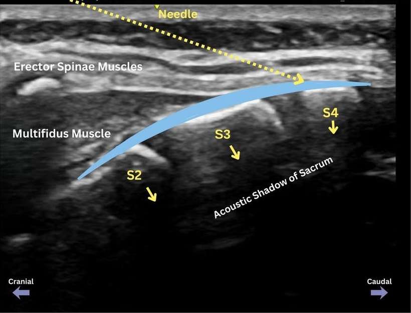
Midline sagittal view over the median crests S1 to S4. The needle comes from above onto S4; one injection spreads to both sides.
- S1–S4
- median sacral crests (fused spinous processes), getting deeper towards the tail
- ESA
- erector spinae aponeurosis over the crests
- Multifidus
- thin multifidus and soft tissue in the midline
- Needle
- in plane from cranial to caudal, onto the S4 crest

Paramedian approach, transverse view. The local anaesthetic (blue) spreads under multifidus, on the bone between the median and intermediate sacral crests.
- Yellow dots
- Needle path (added by the authors)
- Blue
- Spread of local anaesthetic (added by the authors)
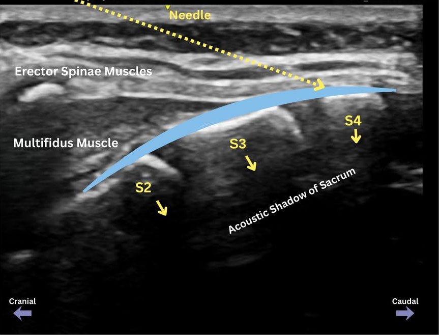
Midline approach, sagittal view. The needle comes in plane from the head end towards S4. The local anaesthetic (blue) spreads under the erector spinae aponeurosis over S2–S4.
- Yellow dots
- Needle path (added by the authors)
- Blue
- Spread of local anaesthetic (added by the authors)
Positioning
Prone or lateral. Operator on the side of the needle; screen opposite.
Scanning technique
- Find the sacral hiatus and cornua, then slide up in the midline to the median sacral crest.
- Midline approach: turn the probe longitudinal (sagittal) over the median crests S1–S4 and insert the needle in plane from above (cranial to caudal) until it touches the S4 crest. Inject deep to the erector spinae aponeurosis; one injection spreads to both sides and may pool towards the sacral hiatus. This approach works best in younger children.
- Paramedian approach: turn the probe transverse at S2, find the intermediate crest about 1.5–2 cm from the midline, and insert the needle in plane from lateral (or from below with a longitudinal probe) onto the crest.
- Watch the drug lift multifidus and erector spinae off the bone and spread up and down the sacrum.
Structures to identify
- Median sacral crest
- Intermediate sacral crest
- Dorsal sacral foramina (gaps in the bone line)
- Multifidus
- Erector spinae aponeurosis and thoracolumbar fascia
- Posterior superior iliac spine
Block performance

Paramedian approach: transverse probe over the sacrum, needle in plane.

Midline approach: probe along the midline of the sacrum, needle in plane from the head end (cranial to caudal).
Position / ergonomics
Stand beside the patient at the level of the pelvis, screen on the other side.
Probe
Linear high-frequency; curvilinear in larger adults.
Settings
Musculoskeletal preset, depth 3–6 cm.
Depth
1–2 cm in children; 3–5 cm in adults.
Needle size
22 G, 50–80 mm.
Needling technique
Paramedian: in plane from lateral (transverse probe) or along the crest (parasagittal probe). Midline: in plane from cranial to caudal onto the S4 crest.
Optimal needle-tip position
On the bone of the median or intermediate crest, deep to multifidus. Not into a dorsal foramen.
Drug choice
See above.
Volume
0.5–1 mL/kg in children; 20–30 mL in adults.
Optimal current if nerve stimulation is used
Not used.
Muscle responses acceptable
Not applicable.
Testing block success
- Loss of cold sensation over the sacrum, perineum or buttock (awake patients)
- No heart rate or blood pressure response to skin incision
Continuous techniques
Not usually used. Catheters have been placed in this plane in a few case reports.
Contraindications to the block
Absolute
- Patient refusal
- Infection over the sacrum (including an infected pilonidal sinus at the needle site)
- Allergy to the drugs used
Relative
- Sacral anomalies such as spina bifida or sacral agenesis
- Coagulopathy or anticoagulants (drug can reach the sacral foramina and epidural space: follow the deep block guidance)
- Local anaesthetic dose limits in small children for bilateral blocks
Side effects and complications
Side effects
- Numb buttock and perineum
- Possible temporary leg weakness or urinary retention if the drug spreads to the sacral roots or epidural space (uncommon)
Complications
- Local anaesthetic toxicity (large volumes, especially bilateral in children)
- Injection into a dorsal foramen or the sacral canal
- Bruising or infection at the site (rare)
Pearls of wisdom
- Start at the sacral hiatus and count upwards: the PSIS line marks S2.
- Keep the needle tip on bone over a crest, not over a gap in the bone line (a foramen).
- For a midline operation, give the volume in the midline or on both sides.
- In children it is an option when parents or surgeons wish to avoid leg weakness after a caudal block.
Review of literature
- Dost, Narayanan and colleagues (2026) reviewed the anatomy, techniques and 14 randomised trials. The block reduced pain and opioid use in adults, and lengthened analgesia compared with caudal or penile blocks in most children's trials. The evidence is still small and varied.
- Rinaldi and Garo (2026) pooled 14 trials (1041 patients): fewer patients needed rescue analgesia and the first rescue came about 5 hours later, but the certainty of evidence was low.
- Kozanhan and colleagues (2026) pooled six children's trials (437 children): longer time to first rescue analgesia and less paracetamol at 24 hours.
- Nair and colleagues (2025) pooled 12 trials of the sacral multifidus plane block (768 patients) and found longer analgesia and less opioid use, with no serious complications.
- Marrone and colleagues (2025) showed on CT spread from S1 up to L2 and to the S2–S3 roots after a single-sided injection.
Comparison with other blocks
| Block | Coverage | Leg weakness | Notes |
|---|---|---|---|
| Sacral ESP / multifidus plane | Sacral dermatomes, perineum; variable lumbar | Rare | Plane block on bone; evidence growing |
| Caudal epidural | Sacral and lower lumbar | Possible | Well established in children |
| Pudendal | Perineum, anus, genitals | Possible (sciatic spread) | Deeper; one nerve each side |
| Dorsal penile | Penis only | None | Circumcision and distal hypospadias |
References
- Dost B, Narayanan M, Marrone F, et al. The sacral erector spinae plane block: a narrative review. Pain Ther 2026;15:131–147. PubMed
- Rinaldi P, Garo ML. Efficacy and safety of sacral erector spinae plane (S-ESP) block in adult and pediatric patients during surgical procedures: a comprehensive systematic review and meta-analysis. J Clin Anesth 2026;111:112181. PubMed
- Kozanhan B, Satici MH, Yildiz M, Tutar MS. Analgesic efficacy of sacral erector spinae plane block in pediatric infraumbilical surgeries: a systematic review and meta-analysis of randomized controlled trials. Minerva Anestesiol 2026;92:209–220. PubMed
- Nair AS, Mistry T, Garg R. Ultrasound-guided sacral multifidus plane block for perioperative analgesia: a comprehensive systematic review, meta-analysis, and trial sequential analysis. Indian J Anaesth 2025;69:1341–1358. PubMed
- Marrone F, Sorrentino T, Megna B, Fusco P, Diwan S. Sacral erector spinae plane block: computed tomography imaging in a hip fracture patient. A A Pract 2025;19:e02105. PubMed
Acknowledgement
Figures 1, 2, 3A, 3B, 4A and 4B on this page are from Dost B, Narayanan M, Marrone F, De Cassai A, Tulgar S, Aksu C, Merjavy P, Chin KJ. The sacral erector spinae plane block: a narrative review. Pain Ther 2026;15:131–147. © The Author(s) 2025. Used under CC BY-NC 4.0. No changes made, except that panels A and B of Figures 3 and 4 are shown separately, and 3B and 4B also appear behind the schematic illustrations.
Test yourself: Sacral erector spinae plane block (sacral multifidus plane block)
4 questions on this page and up to 4 on the picture: name the structure at the numbered dot. Score 70% or more to add this topic to My learning and your certificate. You can try again as often as you like.

New to ultrasound? Start with the Basics of ultrasound or try the scan explorer.