Level of difficulty
Intermediate. Each injection is easy, but there are six on each side, the total dose must be watched, and the superficial temporal artery and the facial nerve branches are close.
Anatomy
The scalp is supplied by branches of the trigeminal nerve at the front and side, and by the cervical nerves at the back.
Front (V1): the supraorbital and supratrochlear nerves, leaving the orbit at the upper rim.
Temple (V2): the zygomaticotemporal nerve, coming out through temporalis behind the lateral orbital rim.
Side (V3): the auriculotemporal nerve, which rises just in front of the ear with the superficial temporal artery.
Back (C2–C3): the greater occipital nerve (C2), which comes up beside the occipital artery about a third of the way from the occipital protuberance to the mastoid; the lesser occipital nerve (C2–C3), further out towards the mastoid; and the third occipital nerve near the midline.
All these nerves run in the dense layer of the scalp above the galea, so small volumes spread well.
Surface anatomy
- Supraorbital notch (in line with the pupil) and the supratrochlear point 1 cm nearer the midline.
- Lateral orbital rim and the zygomatic arch (zygomaticotemporal nerve).
- Tragus of the ear and the superficial temporal artery pulse (auriculotemporal nerve).
- External occipital protuberance, mastoid process and the occipital artery pulse (greater and lesser occipital nerves).
Indications
- Craniotomy: analgesia during and after surgery (PROSPECT recommends a scalp block or incision-site infiltration).
- Awake craniotomy, together with sedation.
- Skull pin fixation (blunts the rise in blood pressure).
- Scalp surgery, lacerations and skin grafts.
- Headache treatment (occipital nerve block, pain clinic).
Nerves blocked
- Supraorbital and supratrochlear (V1)
- Zygomaticotemporal (V2)
- Auriculotemporal (V3)
- Greater occipital (C2)
- Lesser occipital (C2–C3)
- Often the third occipital nerve and the great auricular nerve (C2–C3)
Landmark technique
Surface anatomy, relevant structures and landmarks
Mark each point on both sides before starting, and add up the planned total dose.
Positioning
Supine for the front and side nerves; head turned or the patient on their side for the occipital nerves. In the anaesthetised patient it is often done before pin fixation.
Block technique
- Supraorbital and supratrochlear: 2 mL at the notch and 1–2 mL 1 cm nearer the midline (see the supraorbital page).
- Zygomaticotemporal: 2–3 mL just behind the lateral orbital rim at the level of the upper edge of the zygoma, spread both superficial and deep to temporalis fascia.
- Auriculotemporal: 2–3 mL about 1–1.5 cm in front of the ear at the level of the tragus, after feeling for the superficial temporal artery and injecting beside it (not into it).
- Greater occipital: 3–5 mL beside the occipital artery, about a third of the way along a line from the occipital protuberance to the mastoid.
- Lesser occipital: 2–3 mL, a further 2.5 cm along the same line towards the mastoid.
- Aspirate before each injection and press each site afterwards.
Needle choice
25 G, 25–50 mm.
Neurostimulation technique
Not used.
Choice of local anaesthetic (including volume)
- 0.25–0.5% bupivacaine or levobupivacaine, or 0.5% ropivacaine, often with adrenaline 1:200,000 to slow absorption.
- About 2–5 mL per nerve; a full bilateral block uses 20–30 mL. Check the total dose against the patient's weight: use the dose calculator.
Ultrasound technique
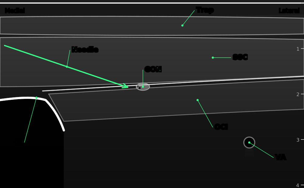
Greater occipital nerve at C2. Transverse view over the C2 spinous process, moved out to the side: the nerve lies between semispinalis capitis and obliquus capitis inferior.
- Trap
- trapezius
- SSC
- semispinalis capitis
- GON
- greater occipital nerve, a small oval on top of obliquus capitis inferior
- OCI
- obliquus capitis inferior, running from C2 out to C1
- C2
- C2 spinous process and lamina with shadow
- VA
- vertebral artery, deep and lateral: keep away
- Needle
- in plane from medial to lateral
Positioning
Sitting, prone or on the side, with the neck slightly bent.
Scanning technique
- Most scalp nerves are blocked by landmarks. Ultrasound is useful mainly for the greater occipital nerve and for finding the arteries.
- Greater occipital at C2: place the probe across the back of the neck over the C2 spinous process (the first large one below the skull), then slide out to the side.
- Tilt the outer end of the probe up along the obliquus capitis inferior muscle. The nerve is the small oval between this muscle and semispinalis capitis above it.
- For the front and side nerves, colour Doppler finds the supraorbital and superficial temporal arteries.
Structures to identify
- Trapezius and semispinalis capitis
- Obliquus capitis inferior
- Greater occipital nerve
- C2 spinous process and lamina
- Vertebral artery (deep, keep away)
Block performance
Position / ergonomics
Move round the head; mark all points first.
Probe
Linear high-frequency, if used.
Settings
Small parts preset, depth 3–4 cm for the occipital nerve.
Depth
0.3–1 cm for the front and side nerves; about 1.5–2 cm for the greater occipital nerve at C2.
Needle size
25 G, 25–50 mm.
Needling technique
Landmark for most; in plane from medial to lateral for the greater occipital nerve at C2.
Optimal needle-tip position
Next to each nerve, outside any artery. At C2, in the plane between semispinalis capitis and obliquus capitis inferior.
Drug choice
See above.
Volume
2–5 mL per nerve.
Optimal current if nerve stimulation is used
Not used.
Muscle responses acceptable
Not applicable.
Testing block success
- Numb scalp in the area of each nerve (awake patients)
- Little rise in blood pressure or heart rate at pin fixation and skin incision
Continuous techniques
Not usually used. The block can be repeated at the end of surgery if it was given before pin fixation.
Contraindications to the block
Absolute
- Patient refusal
- Infection at the injection sites
- Allergy to the drugs used
Relative
- Skull defect or fracture at an injection site
- Coagulopathy (the injections are superficial and compressible)
- Local anaesthetic dose limits in small patients (many injections, large total volume)
Side effects and complications
Side effects
- Bruising and swelling, especially round the eyes
- Numb scalp for several hours
Complications
- Intravascular injection (superficial temporal or occipital artery)
- Local anaesthetic toxicity from the large total dose
- Temporary facial nerve weakness if the auriculotemporal injection is too deep
- Injection into the orbit (supraorbital)
Pearls of wisdom
- Work out the total dose before you start: twelve injections add up quickly.
- Feel the superficial temporal artery and inject beside it, staying shallow to avoid the facial nerve.
- Doing the block before pin fixation blunts the blood pressure surge.
- The surgeon can add infiltration along the incision, but count it in the total dose.
Review of literature
- Guilfoyle and colleagues (2013) pooled seven trials (320 patients): scalp block reduced pain after craniotomy and opioid use over 24 hours, with no complications reported.
- Duda and colleagues (2023) updated the evidence in a larger meta-analysis and found less pain and less opioid use after scalp block for craniotomy.
- The PROSPECT group (Mestdagh 2023) recommends a scalp block or incision-site infiltration for elective craniotomy, as part of multimodal analgesia.
- Osborn and Sebeo (2010) describe the anatomy, history and technique of the scalp block.
Comparison with other blocks
| Technique | Coverage | Total dose | Notes |
|---|---|---|---|
| Full scalp block | Whole scalp, both sides | High | Craniotomy, awake craniotomy, pins |
| Incision-site infiltration | Wound only | Lower | Simple; recommended alternative |
| Selective nerve blocks | Part of the scalp | Low | Small incisions, headache treatment |
References
- Guilfoyle MR, Helmy A, Duane D, Hutchinson PJA. Regional scalp block for postcraniotomy analgesia: a systematic review and meta-analysis. Anesth Analg 2013;116:1093–1102. PubMed
- Duda T, Lannon M, Gandhi P, et al. Systematic review and meta-analysis of randomized controlled trials for scalp block in craniotomy. Neurosurgery 2023;93:4–23. PubMed
- Mestdagh FP, Lavand'homme PM, Pirard G, et al. Pain management after elective craniotomy: a systematic review with procedure-specific postoperative pain management (PROSPECT) recommendations. Eur J Anaesthesiol 2023;40:747–757. PubMed
- Osborn I, Sebeo J. 'Scalp block' during craniotomy: a classic technique revisited. J Neurosurg Anesthesiol 2010;22:187–194. PubMed
Test yourself: Scalp block
4 questions on this page and up to 4 on the picture: name the structure at the numbered dot. Score 70% or more to add this topic to My learning and your certificate. You can try again as often as you like.

New to ultrasound? Start with the Basics of ultrasound or try the scan explorer. Compare this block with another.