Level of difficulty
Basic. The target is shallow and easy to see, and there are no large structures in the needle path when the needle stays superficial to the investing fascia.
Anatomy
The cervical plexus is formed by the front branches of C1–C4. Its four sensory (cutaneous) branches wind round the back edge of sternocleidomastoid at about its midpoint, at a spot often called Erb's point (or the nerve point of the neck).
The four branches are the lesser occipital (behind the ear), great auricular (ear lobe and skin over the angle of the jaw), transverse cervical (front of the neck) and supraclavicular nerves (skin over the collarbone and top of the shoulder).
They emerge between the investing layer of deep cervical fascia and the platysma. A superficial block puts the drug just under the platysma here, above the investing fascia.
The external jugular vein crosses sternocleidomastoid at about the same level, and is a useful landmark and a structure to avoid.
Surface anatomy
- Mastoid process.
- Clavicular head of sternocleidomastoid.
- Midpoint of the back edge of sternocleidomastoid (about the level of the cricoid cartilage, C6, or a little higher at C4).
- External jugular vein crossing the muscle.
Indications
- Thyroid and parathyroid surgery (usually bilateral), as part of multimodal analgesia.
- Carotid endarterectomy (with surgical infiltration), as the regional anaesthetic for awake surgery.
- Superficial neck surgery: lymph node biopsy, skin lesions, tracheostomy (bilateral).
- Clavicle fracture surgery, together with an interscalene or superior trunk block.
- Ear and mastoid skin (great auricular nerve), tympanomastoid surgery in children.
Nerves blocked
- Lesser occipital nerve (C2)
- Great auricular nerve (C2–C3)
- Transverse cervical nerve (C2–C3)
- Supraclavicular nerves (C3–C4)
Landmark technique
Surface anatomy, relevant structures and landmarks
Draw a line from the mastoid to the clavicular head of sternocleidomastoid and mark its midpoint on the back edge of the muscle.
Positioning
Supine with the head turned about 30° away from the side to be blocked. Ask the patient to lift the head slightly to make the muscle stand out.
Block technique
- Insert the needle just under the skin at the marked point, behind the back edge of the muscle.
- Inject 3–5 mL, then redirect the needle up and down along the back edge of the muscle (a 'fan'), injecting a further 2–3 mL in each direction, staying just under the skin.
- Aspirate before each injection and avoid the external jugular vein.
Needle choice
25 G or 23 G hypodermic needle, or a 22 G short-bevel 50 mm needle.
Neurostimulation technique
Not used: the branches are purely sensory.
Choice of local anaesthetic (including volume)
- 5–10 mL each side of 0.25–0.5% levobupivacaine or bupivacaine, or 0.375–0.5% ropivacaine.
- For bilateral blocks keep the total dose within the maximum: use the dose calculator.
Ultrasound technique
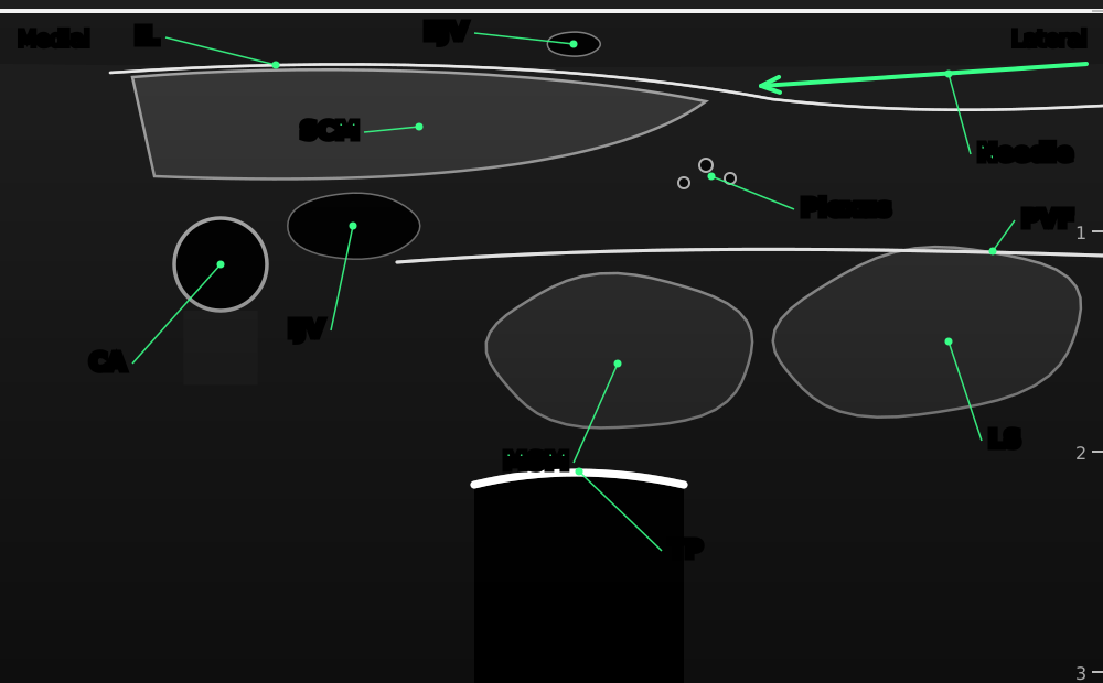

Transverse view at the middle of the back edge of sternocleidomastoid. The cervical plexus branches lie as small dark dots just behind and under the edge of the muscle, superficial to the prevertebral fascia.
- SCM
- sternocleidomastoid, tapering at its posterior (lateral) border
- IL
- investing layer of the deep cervical fascia: the white layer over sternocleidomastoid
- EJV
- external jugular vein on the surface of SCM
- Plexus
- small branches of the superficial cervical plexus near the nerve point
- CA
- common carotid artery
- IJV
- internal jugular vein
- PVF
- prevertebral fascia (keep superficial to it)
- MSM
- middle scalene muscle
- LS
- levator scapulae
- TP
- C4 transverse process with acoustic shadow
- Needle
- in plane from lateral, staying just under the skin
Positioning
As for the landmark technique, with the operator at the head end or on the side being blocked.
Scanning technique
- Place the probe across the neck at the level of the cricoid, over sternocleidomastoid.
- Slide laterally until the tapered back edge of the muscle is in the middle of the screen.
- Look for the small cluster of hypoechoic nerve branches just behind the edge of the muscle, on top of the levator scapulae and scalene muscles.
- Use colour Doppler to find the external jugular vein and small arteries.
Structures to identify
- Sternocleidomastoid
- Investing fascia and platysma
- Cervical plexus branches ('nerve point')
- External jugular vein
- Prevertebral fascia over levator scapulae and the scalenes
- Carotid artery and internal jugular vein (medial, deep: keep away)
Block performance

Position / ergonomics
Stand at the head of the bed or on the side of the block, with the screen in line with the needle.
Probe
Linear, high frequency (10–15 MHz).
Settings
Small parts or nerve preset, depth 2–3 cm.
Depth
0.5–1.5 cm.
Needle size
22–25 G, 50 mm short-bevel.
Needling technique
In-plane from lateral to medial (or out of plane), keeping the tip shallow.
Optimal needle-tip position
Just under the platysma beside the back edge of sternocleidomastoid, superficial to the investing fascia. Local anaesthetic should spread under the skin along the edge of the muscle.
Drug choice
Long-acting local anaesthetic, as above.
Volume
5–10 mL each side.
Optimal current if nerve stimulation is used
Not used.
Muscle responses acceptable
Not applicable (sensory nerves).
Testing block success
- Loss of cold and pinprick sensation over the front and side of the neck
- Numb ear lobe (great auricular nerve)
- Numb skin over the clavicle
Continuous techniques
Not usually used. A single injection covers the first postoperative day for most neck surgery.
Contraindications to the block
Absolute
- Patient refusal
- Infection at the injection site
- Allergy to the drugs used
Relative
- Coagulopathy (a superficial, compressible block, so often acceptable after weighing the risks)
- Distorted neck anatomy (previous surgery, radiotherapy, large goitre)
- Contralateral phrenic or recurrent laryngeal nerve palsy (avoid bilateral deeper spread)
Side effects and complications
Side effects
- Numb neck and ear lobe for several hours
- Bruising
Complications
- Intravascular injection (external jugular vein)
- Spread deeper than intended, with phrenic nerve, recurrent laryngeal nerve or Horner's syndrome (rare with a truly superficial injection)
- Local anaesthetic toxicity with large bilateral volumes
- Infection (rare)
Pearls of wisdom
- Stay superficial: the safety of this block comes from staying above the investing fascia.
- It numbs only the skin. For carotid surgery the surgeon will usually need to add infiltration around the carotid sheath and the jaw.
- For thyroid surgery give it on both sides before the incision; benefit is modest but consistent.
- Scan with colour Doppler first: the external jugular vein is often right over the target.
Review of literature
- Pandit and colleagues (2007) reviewed over 10,000 blocks for carotid endarterectomy. Superficial or intermediate blocks had fewer needle-related complications and fewer conversions to general anaesthesia than deep or combined blocks.
- Wilson and colleagues (2023) pooled 31 trials (2,273 patients) of bilateral superficial cervical plexus block for thyroid surgery: less opioid use, lower pain scores to 24 h and less sickness.
- Kim and colleagues (2018) reviewed the anatomy of the cervical fascial layers and proposed naming cervical plexus blocks by the compartment where the drug goes.
Comparison with other blocks
| Block | Injection site | Phrenic palsy risk | Main use |
|---|---|---|---|
| Superficial cervical plexus | Under platysma, above investing fascia | Very low | Thyroid, superficial neck, clavicle skin |
| Intermediate cervical plexus | Between SCM and prevertebral fascia | Low | Carotid endarterectomy, neck surgery |
| Deep cervical plexus | At the C2–C4 transverse processes | High | Rarely used now |
References
- Pandit JJ, Satya-Krishna R, Gration P. Superficial or deep cervical plexus block for carotid endarterectomy: a systematic review of complications. Br J Anaesth 2007;99:159–169. PubMed
- Wilson L, Malhotra R, Mayhew D, Banerjee A. The analgesic effects of bilateral superficial cervical plexus block in thyroid surgery: a systematic review and meta-analysis. Indian J Anaesth 2023;67:579–589. PubMed
- Kim JS, Ko JS, Bang S, Kim H, Lee SY. Cervical plexus block. Korean J Anesthesiol 2018;71:274–288. PubMed
Test yourself: Superficial cervical plexus block
4 questions on this page and up to 4 on the scan: name the structure at the numbered dot. Score 70% or more to add this topic to My learning and your certificate. You can try again as often as you like.
New to ultrasound? Start with the Basics of ultrasound or try the scan explorer. Compare this block with another.