Level of difficulty
Basic. The notch can usually be felt, and the injection is shallow.
Anatomy
The supraorbital and supratrochlear nerves are end branches of the frontal nerve, from the ophthalmic division of the trigeminal nerve (V1).
The supraorbital nerve leaves the orbit through the supraorbital notch or foramen on the upper orbital rim, about 2–2.5 cm from the midline, in line with the pupil when the eye looks straight ahead. It runs up under frontalis to supply the forehead and scalp as far back as the crown.
The supratrochlear nerve leaves the orbit about 1 cm nearer the midline, above the trochlea, and supplies the middle of the forehead and the upper eyelid.
The supraorbital artery runs with the nerve and can be seen with colour Doppler.
Surface anatomy
- Supraorbital notch: on the upper orbital rim in line with the pupil.
- Supratrochlear point: about 1 cm towards the midline from the notch, at the upper inner corner of the orbit.
Indications
- Forehead and front-scalp skin surgery, lacerations and skin grafts.
- Part of a scalp block for craniotomy, including awake craniotomy and pin fixation.
- Upper eyelid and brow surgery.
- Headache and supraorbital neuralgia (pain clinic).
Nerves blocked
- Supraorbital nerve (V1)
- Supratrochlear nerve (V1)
Landmark technique
Surface anatomy, relevant structures and landmarks
Feel the notch on the upper orbital rim in line with the pupil.
Positioning
Supine, eyes closed. Put a finger on the upper orbital rim to keep the needle out of the orbit.
Block technique
- Insert the needle just above the notch, aiming slightly upwards, until it touches bone or lies just above the notch. Do not enter the foramen.
- Aspirate and inject 1–2 mL.
- For the supratrochlear nerve, move about 1 cm towards the midline and inject 1 mL; or make a line of injection along the brow from the notch to the midline.
- Press firmly for a minute afterwards to limit bruising.
Needle choice
25–27 G, 25 mm.
Neurostimulation technique
Not used.
Choice of local anaesthetic (including volume)
- 1–2 mL per nerve of 1–2% lidocaine with adrenaline, or 0.25–0.5% levobupivacaine or bupivacaine for longer effect.
Ultrasound technique
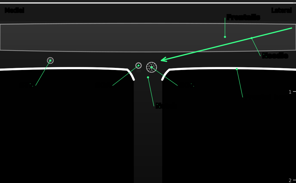

Transverse view along the upper orbital rim. The bright line of the frontal bone has a gap at the supraorbital notch, where the nerve and artery leave the orbit under frontalis.
- Frontalis
- frontalis and orbicularis oculi muscles
- Frontal bone
- upper orbital rim (bright line with shadow)
- Notch
- supraorbital notch or foramen: a gap in the bone line
- SON
- supraorbital nerve
- SOA
- supraorbital artery (colour Doppler)
- STN
- supratrochlear nerve, about 1 cm nearer the midline
- Needle
- in plane from the side (out of plane also works)
Positioning
Supine, with the operator at the head end.
Scanning technique
- Place a small linear probe along the brow, just above the upper orbital rim.
- Slide along the rim to find the break in the bright bone line: the notch or foramen.
- Colour Doppler shows the supraorbital artery in the notch.
Structures to identify
- Frontal bone (bright line)
- Supraorbital notch or foramen
- Supraorbital artery
- Supraorbital nerve
- Frontalis and orbicularis oculi
Block performance
Position / ergonomics
Stand at the head of the bed.
Probe
Small linear high-frequency (hockey-stick) probe.
Settings
Superficial or small parts preset, depth 1.5–2 cm.
Depth
0.3–0.8 cm.
Needle size
25–27 G, 25 mm.
Needling technique
Out of plane, or in plane from the side, aiming at the notch.
Optimal needle-tip position
Next to the notch, superficial to the bone and outside the foramen.
Drug choice
See above.
Volume
1–2 mL per nerve.
Optimal current if nerve stimulation is used
Not used.
Muscle responses acceptable
Not applicable.
Testing block success
- Numb forehead and front of the scalp on that side
Continuous techniques
Not used.
Contraindications to the block
Absolute
- Patient refusal
- Infection at the injection site
- Allergy to the drugs used
Relative
- Fracture of the orbital rim or frontal bone at the site
- Coagulopathy (usually acceptable: superficial and compressible)
Side effects and complications
Side effects
- Bruising and swelling of the upper eyelid (common)
- Numb forehead
Complications
- Injection into the orbit (keep a finger on the rim)
- Intravascular injection
- Nerve injury from injecting into the foramen (rare)
Pearls of wisdom
- Keep a finger on the orbital rim so the needle cannot slip into the orbit.
- Press on the site after injection; the eyelid bruises easily.
- Block both nerves: the supratrochlear covers the middle of the forehead.
- For a midline incision, block both sides.
Review of literature
- Allam and colleagues (2018) reviewed the anatomy and ultrasound-guided techniques for the supraorbital, infraorbital, mental and other trigeminal branches.
- Osborn and Sebeo (2010) described the supraorbital and supratrochlear blocks as the front part of the scalp block for craniotomy.
- Guilfoyle and colleagues (2013) pooled seven trials of scalp blocks (including these nerves) for craniotomy and found less pain in the first hours after surgery.
Comparison with other blocks
| Block | Area numbed | Division |
|---|---|---|
| Supraorbital and supratrochlear | Forehead, front of the scalp, upper eyelid | V1 |
| Infraorbital | Lower eyelid, cheek, side of nose, upper lip | V2 |
| Mental | Lower lip and chin | V3 |
| Scalp block | Whole scalp | V1–V3 and C2–C3 |
References
- Allam AE, Khalil AAF, Eltawab BA, Wu WT, Chang KV. Ultrasound-guided intervention for treatment of trigeminal neuralgia: an updated review of anatomy and techniques. Pain Res Manag 2018;2018:5480728. PubMed
- Osborn I, Sebeo J. 'Scalp block' during craniotomy: a classic technique revisited. J Neurosurg Anesthesiol 2010;22:187–194. PubMed
- Guilfoyle MR, Helmy A, Duane D, Hutchinson PJA. Regional scalp block for postcraniotomy analgesia: a systematic review and meta-analysis. Anesth Analg 2013;116:1093–1102. PubMed
Test yourself: Supraorbital and supratrochlear nerve block
4 questions on this page and up to 3 on the scan: name the structure at the numbered dot. Score 70% or more to add this topic to My learning and your certificate. You can try again as often as you like.
New to ultrasound? Start with the Basics of ultrasound or try the scan explorer. Compare this block with another.