FoCUS · SHOCK · BEFORE SURGERY
Focused cardiac ultrasound
When to use it
- Find the cause of low blood pressure or shock: an empty heart (hypovolaemia), a weak heart, a dilated right ventricle (for example, PE), or fluid around the heart (tamponade).
- Before surgery, look at a patient with a new murmur or unexplained breathlessness (for example, a stiff, calcified aortic valve). Refer for formal echocardiography when in doubt.
- During cardiac arrest, look for treatable causes, in pauses of under 10 seconds.
- Probe
- Phased array (cardiac), low frequency
- Position
- Supine or left lateral; cardiac preset
- Depth
- 14–18 cm
How to scan
- Parasternal long axis: left of the sternum, 3rd–4th space, probe marker towards the right shoulder.
- Parasternal short axis: turn the probe 90° clockwise. Look for a round left ventricle and a crescent-shaped right ventricle.
- Apical four-chamber: at the apex beat, marker towards the left. Compare the size of the right and left ventricles.
- Subcostal four-chamber and IVC: under the xiphoid, aimed at the left shoulder (good when ventilated or in arrest). Turn to see the IVC entering the right atrium.
- Answer simple questions: Is the LV squeezing well? Is the RV big? Is there fluid around the heart? Is the heart empty and the IVC small?
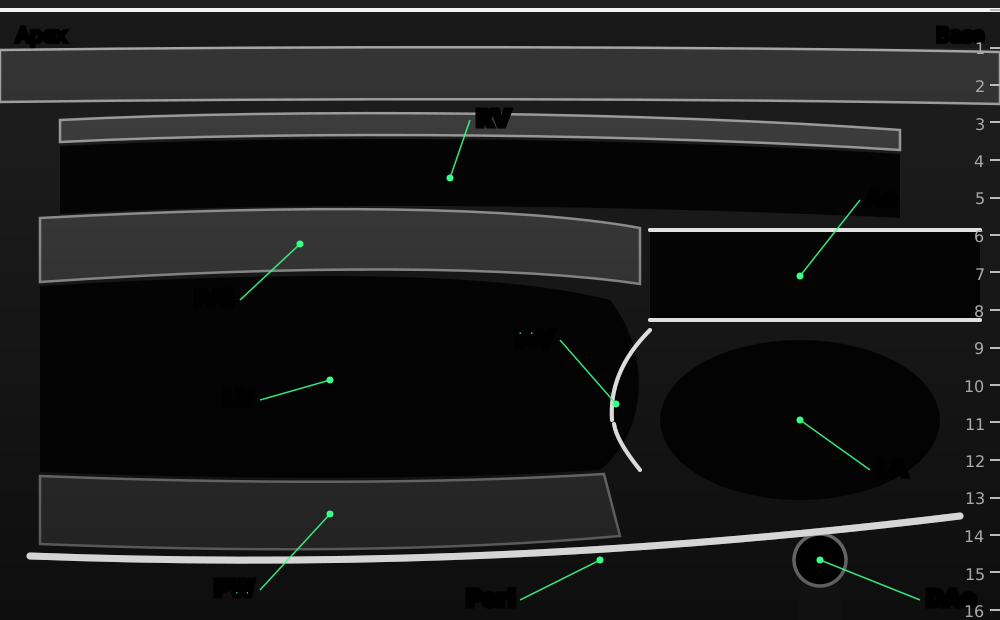
Parasternal long-axis view: right ventricle at the top, then septum, left ventricle and posterior wall; aortic root and left atrium on the right.
- RV
- right ventricle, at the top of the screen
- IVS
- interventricular septum
- LV
- left ventricle
- PW
- posterior wall of the left ventricle
- Ao
- aortic root and valve
- LA
- left atrium
- MV
- mitral valve
- Peri
- pericardium: fluid shows as a black stripe between it and the heart
- DAo
- descending aorta (an effusion lies in front of it, a pleural effusion behind it)
Original schematic illustration © Nerve Blocks. Not to scale.
What you see and what it means
| On the screen | What it means |
|---|---|
| Small, vigorous LV; walls touch in systole; small, collapsing IVC | Hypovolaemia: give fluid |
| Big LV that barely moves | Poor LV function |
| RV as big as or bigger than LV; flat septum | RV strain: think PE |
| Black stripe around the heart; collapsing RV or RA | Pericardial effusion; tamponade if the patient is in shock |
| Bright, stiff aortic valve that barely opens | Possible severe aortic stenosis: formal echo |
Pearls
- FoCUS answers yes/no questions. It is not a full echocardiogram: refer when the answer matters and you are unsure.
- Look at more than one view before deciding. One view can mislead.
- Pericardial fluid lies in front of the descending aorta, pleural fluid behind it.
References
- Via G, Hussain A, Wells M, et al. International evidence-based recommendations for focused cardiac ultrasound. J Am Soc Echocardiogr 2014;27:683.e1–683.e33. PubMed
