The routine for every block

Follow the same steps every time. 'Stop Before You Block' comes just before the needle touches the skin.
Original diagram © Nerve Blocks.
Preparation and planning
- Patient: history, allergies, anticoagulants and antiplatelets (see our anticoagulation guide), lung function for blocks that may affect the phrenic nerve, existing nerve problems (document them before the block).
- Block choice: match the block to the operation (try our block finder), how long you need it to last, and the patient's other conditions.
- Consent: benefits, common side effects (numbness, weakness, falls risk), rare but serious risks (nerve injury, toxicity, pneumothorax where relevant), alternatives, and what to expect afterwards.
- Kit: ultrasound machine with the right probe, sterile cover and gel, echogenic needle, nerve stimulator if used, drugs drawn up and labelled, monitoring, oxygen, suction, resuscitation drugs and lipid emulsion 20%.
- Place: a block room or quiet bay with enough space, good light and a trained assistant.
Monitoring and access
- IV access before the block. Standard monitoring (ECG, blood pressure, oxygen saturation) during the block and for at least 30 minutes after, when toxicity is most likely.
- Light sedation only, if any: the patient should be able to tell you about pain or tingling.
Asepsis
- Hand hygiene, sterile gloves; gown, hat and mask for catheters.
- Clean the skin with chlorhexidine in alcohol and let it dry completely: chlorhexidine is toxic to nerves if injected.
- Sterile probe cover and sterile gel. Keep the sterile field intact throughout.
Stop Before You Block
Immediately before inserting the needle, the operator and assistant check the site mark and the side again, together. Wrong-side blocks still happen, often after a delay or a change of position. See our emergency protocols.
Injection
- Aspirate first. Inject 0.5–1 mL and watch: you should see the fluid spread in the right plane.
- Inject in 3–5 mL aliquots, aspirating between them, and keep watching the spread.
- Low pressure only. Stop if it is hard to inject, if the patient feels pain or tingling, or if no spread appears (the tip may be in a vessel).
- Count the total dose of local anaesthetic, including any infiltration by the surgeon: use our dose calculator.
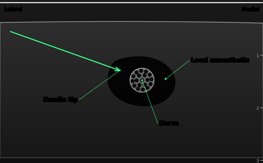
Aim for spread that surrounds the nerve or opens the fascial plane. No visible spread means the tip is not where you think.
Original schematic illustration © Nerve Blocks. Not to scale.
Common pitfalls for beginners
- Advancing or injecting when the tip is not in view.
- Too much probe pressure: veins disappear and the anatomy changes.
- Wrong depth or gain: the target or needle is lost.
- Injecting without seeing where the fluid goes.
- Choosing a block beyond your experience, or the patient's anatomy, without supervision.
After the block
- Test the block before surgery: cold, touch and movement in the expected area. Have a plan if it is incomplete.
- Document: block, side, approach, needle, drug, concentration, volume, any difficulties, the patient's response.
- Protect the numb limb (sling, care with heat, falls precautions after lower limb blocks).
- Prescribe regular painkillers to start before the block wears off, to avoid rebound pain.
- Tell the patient who to call if numbness or weakness lasts longer than expected.
References
- Marhofer P, Chan VWS. Ultrasound-guided regional anesthesia: current concepts and future trends. Anesth Analg 2007;104:1265–9. PubMed
